Review
. 2008 Dec;9(12):1264-76.

HER-2-directed, small-molecule antagonists

Affiliations
Review

HER-2-directed, small-molecule antagonists

Michelle Arkin et al. Curr Opin Investig Drugs. 2008 Dec.

Abstract

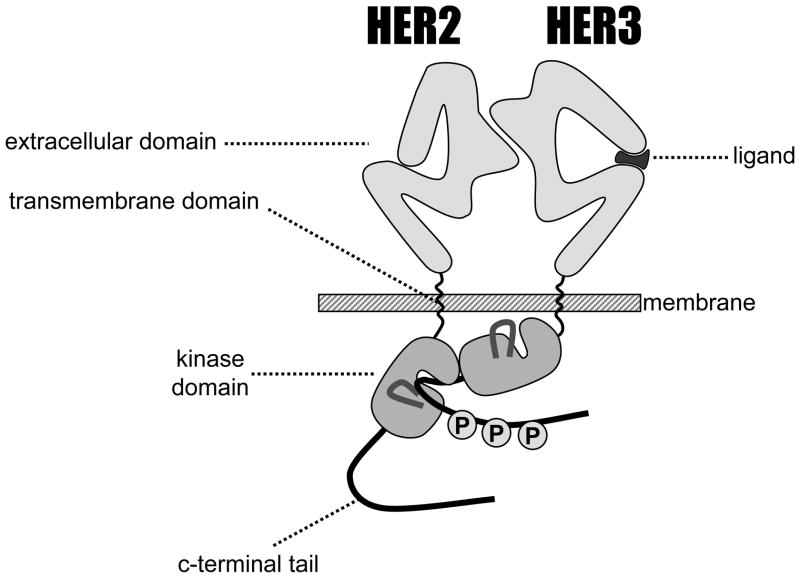
Inactivation of the human epidermal growth factor receptor-2 (HER-2) tyrosine kinase holds significant promise as a cancer treatment hypothesis, making it a high-value target for drug discovery. Screening and structure-based efforts have led to the development of several classes of ATP analog inhibitors of the HER-2 tyrosine kinase. These efforts have been further enhanced by detailed structural information regarding its sibling kinase, the EGF receptor, and structural properties that can be exploited to confer activity and even selectivity toward HER-2 kinase. Signaling and structural studies also suggest a critical involvement of the kinase inactive HER-3 in the regulation of HER-2, creating unique challenges in the efforts to inactivate the latter.


Figures

Figure 1
Figure 1
Schematic representation of the HER2/3 heterodimer.
Figure 2
Figure 2
Schematic representation of HER kinase domain conformations. The kinase domain contains a beta-strand rich N-lobe and a helix-rich C-lobe connected by a hinge region. The ATP and peptide substrates bind in the cleft between the two domains, and ATP forms hydrogen bonds with the backbone of the hinge (see Figure 3). Left, the inactive conformation is characterized by a narrow opening between the N- and C-lobes and occlusion of the active site by the Activation loop. The regulatory alpha-C helix (αC), which contains a critical glutamic acid residue (E738), is oriented away from the active site. Right, the active conformation of the kinase domain has a more open cleft for binding substrates and the activation loop is directed away from the binding site. The conformation of the αC helix allows E738 to make a hydrogen bond with the catalytic lysine residue (K721). Image adapted from [5].
Figure 3
Figure 3
Hydrogen bonding between a nonhydrolyzable ATP analog and the EGFR kinase domain. The adenosine ring of ATP (cyan) forms two hydrogen bonds with the backbone of the hinge region of EGFR kinase domain (green). Tyrosine kinase inhibitors contain an ATP-mimetic core that makes at least one of these hydrogen bonds; some ATP-mimetics also hydrogen bond to the carbonyl of the next residue (proline in EGFR). While the hinge residues vary, the backbone conformation and hydrogen-bonding orientation is common among kinases. Image derived from Protein Databank file 2ITN [33].
Figure 4
Figure 4
Selected HER TKI compounds. Core structures for each compound class are shown in black; functional groups that vary between compounds within a class are shown in gray.
Figure 5
Figure 5
X-ray structures of EGFR bound to erlotinib (left; [29]) and lapatinib (right; [17]). The erlotinib-bound protein adopts the active conformation, with a wide cleft between the N- and C-lobes (blue and green, respectively), and the activation loop (orange) and C-terminus (yellow) oriented away from the active site. By contrast, the lapatinib-bound protein adopts the inactive conformation. Here, the protein surrounds the small molecule, with the N-lobe (blue) rotating towards the binding cleft, and the activation loop (orange) and αC helix (cyan) obstructing the substrate binding site. The C-terminal tail (yellow) also packs against the “left” side of the kinase. The 4-anilino-quinazoline core is oriented similarly in both structures and makes the same hydrogen bond to the methionine backbone in the hinge sequence (dark blue). Images derived from Protein Databank files 1M17 [29] and 1XKK [17].
Figure 6
Figure 6
X-ray structures comparing the orientation of compounds that bind to the active (left) and inactive (right) conformations of EGFR kinase domain. Left: The pyrrolopyrimidine core of AEE788 (pink) overlays closely with the quinazoline core of gefitinib (cyan). The anilino groups also occupy a similar pocket in the enzyme. Right: The cyanoquinoline core of HKI-272 (green) overlays closely with the quinazoline core of lapatinib (blue). The anilino groups also align. HKI-272 covalently modifies EGFR at Cys797, shown in yellow. The main chain atoms that hydrogen-bond to the inhibitors are shown as spheres. Images derived from Protein Databank files 2ITY [33], 2J6M [33], 1XKK [17], and 2JIV [49].

References

    1. Ferguson KM. Structure-based view of epidermal growth factor receptor regulation. Annu Rev Biophys. 2008;37:353–373. - PMC - PubMed
    1. Berger MB, Mendrola JM, Lemmon MA. ErbB3/HER3 does not homodimerize upon neuregulin binding at the cell surface. FEBS Lett. 2004;569(1–3):332–336. - PubMed
    1. Sierke SL, Cheng K, Kim HH, Koland JG. Biochemical characterization of the protein tyrosine kinase homology domain of the ErbB3 (HER3) receptor protein. Biochemical Journal. 1997;322(Pt 3):757–763. - PMC - PubMed
    1. Tzahar E, Waterman H, Chen X, Levkowitz G, Karunagaran D, Lavi S, Ratzkin BJ, Yarden Y. A hierarchical network of interreceptor interactions determines signal transduction by Neu differentiation factor/neuregulin and epidermal growth factor. Molecular and Cellular Biology. 1996;16:5276–5287. - PMC - PubMed
    1. Zhang X, Gureasko J, Shen K, Cole PA, Kuriyan J. An allosteric mechanism for activation of the kinase domain of epidermal growth factor receptor. Cell. 2006;125(6):1137–1149. - PubMed

MeSH terms

LinkOut - more resources