

Review
doi: 10.1016/j.neuron.2008.11.005.
Striatal plasticity and basal ganglia circuit function
Affiliations
- PMID: 19038213
- PMCID: PMC2724179
- DOI: 10.1016/j.neuron.2008.11.005
Item in Clipboard
Review
Striatal plasticity and basal ganglia circuit function
Neuron.
.
Abstract
The dorsal striatum, which consists of the caudate and putamen, is the gateway to the basal ganglia. It receives convergent excitatory afferents from cortex and thalamus and forms the origin of the direct and indirect pathways, which are distinct basal ganglia circuits involved in motor control. It is also a major site of activity-dependent synaptic plasticity. Striatal plasticity alters the transfer of information throughout basal ganglia circuits and may represent a key neural substrate for adaptive motor control and procedural memory. Here, we review current understanding of synaptic plasticity in the striatum and its role in the physiology and pathophysiology of basal ganglia function.
Figures
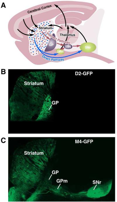
Sagittal view of a mouse brain (A), depicting cortex-basal ganglia-thalamus-cortex circuits. Axons from the thalamus and striatum form excitatory synapses onto striatonigral/direct-pathway MSNs (blue) and striatopallidal/indirect-pathway MSNs (red). Direct pathway MSNs send axons directly to basal ganglia output nuclei (medial globus pallidus, GPm, and substantia nigra, pars reticulata, SNr), where they form inhibitory synapses. Indirect-pathway MSNs inhibit neurons in the globus pallidus (GP), which in turn make inhibitory connections with the subthalamic nucleus (STN). STN projections target the GPm and SNr, where they form excitatory synapses onto GABAergic basal ganglia output neurons. These inhibitory output neurons send axons to ventroposterior thalamic motor nuclei. Finally, glutamatergic neurons in the thalamus project back to cortex, completing the circuit. A sagittal slice from a BAC-transgenic mouse expressing GFP under the control of genomic regulatory elements for the dopamine D2 receptor (D2-GFP) (B), or the muscarinic M4 receptor (M4-GFP) (C), which labels indirect- and direct-pathway MSNs, respectively. Figure inspired by (Gerfen, 2006).
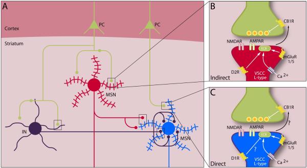
(A) Simplified schematic of striatal neurons and their interconnections. Cortical pyramidal neurons (green) project to striatal interneurons (INTs) and medium spiny neurons (MSNs) of the direct (blue) and indirect (red) pathways. Interneurons also form synapses on medium spiny neurons. Rectangles highlight potential sites of synaptic plasticity that could alter striatal output from MSNs. Corticostriatal synapses on direct and indirect pathway MSNs are expanded at right. (B) Indirect-pathway spines contain dopamine D2 receptors (D2R), group I mGluRs (mGluR1/5), and L-type voltage-sensitive calcium channels (VSCCs), which synergistically mobilize endocannabinoid (eCB) release that can induce presynaptic LTD by acting at cannabinoid receptors (CB1R). (C) Direct-pathway spines contain dopamine D1 receptors (D1R), group I mGluRs, and L-type VSCCs. Endocannabinoid-dependent LTD reportedly occurs at direct-pathway MSNs under conditions in which D1 receptors are not activated.
References
-
- Albin RL, Young AB, Penney JB. The functional anatomy of basal ganglia disorders. Trends Neurosci. 1989;12:366–375. - PubMed
-
- Alexander GE, Crutcher MD. Functional architecture of basal ganglia circuits: neural substrates of parallel processing. Trends Neurosci. 1990;13:266–271. - PubMed
-
- Aouizerate B, Guehl D, Cuny E, Rougier A, Bioulac B, Tignol J, Burbaud P. Pathophysiology of obsessive-compulsive disorder: a necessary link between phenomenology, neuropsychology, imagery and physiology. Prog. Neurobiol. 2004;72:195–221. - PubMed
-
- Aravanis AM, Wang LP, Zhang F, Meltzer LA, Mogri MZ, Schneider MB, Deisseroth K. An optical neural interface: in vivo control of rodent motor cortex with integrated fiberoptic and optogenetic technology. J. Neural Eng. 2007;4:S143–S156. - PubMed
Publication types
MeSH terms
Grants and funding
LinkOut - more resources
Full Text Sources
Other Literature Sources
