. 2008 Nov 26;60(4):610-24.
doi: 10.1016/j.neuron.2008.09.024.

A biological function for the neuronal activity-dependent component of Bdnf transcription in the development of cortical inhibition

Affiliations

A biological function for the neuronal activity-dependent component of Bdnf transcription in the development of cortical inhibition

Elizabeth J Hong et al. Neuron. .

Abstract

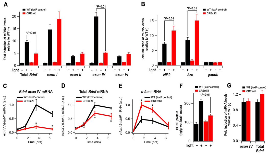
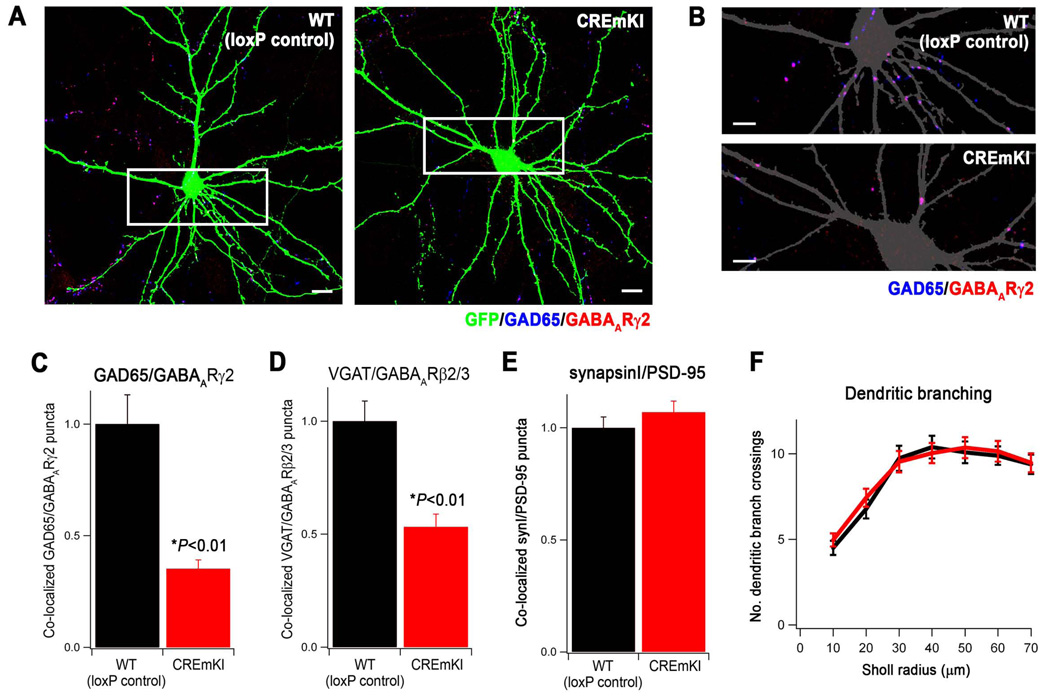
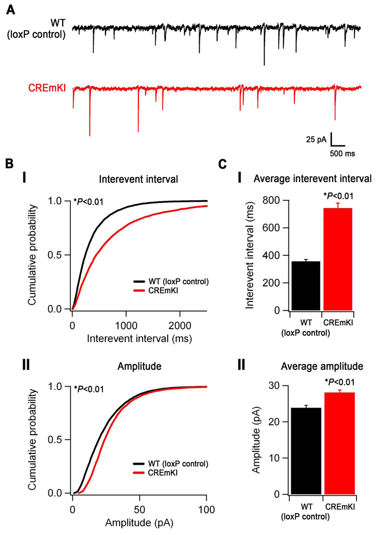
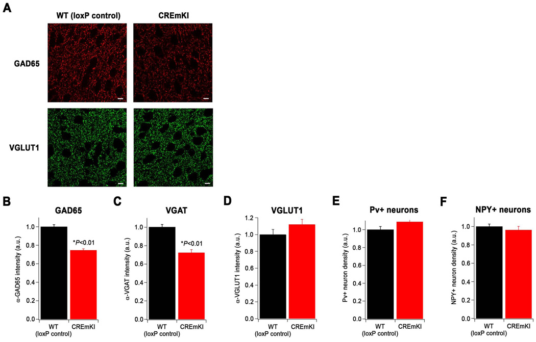
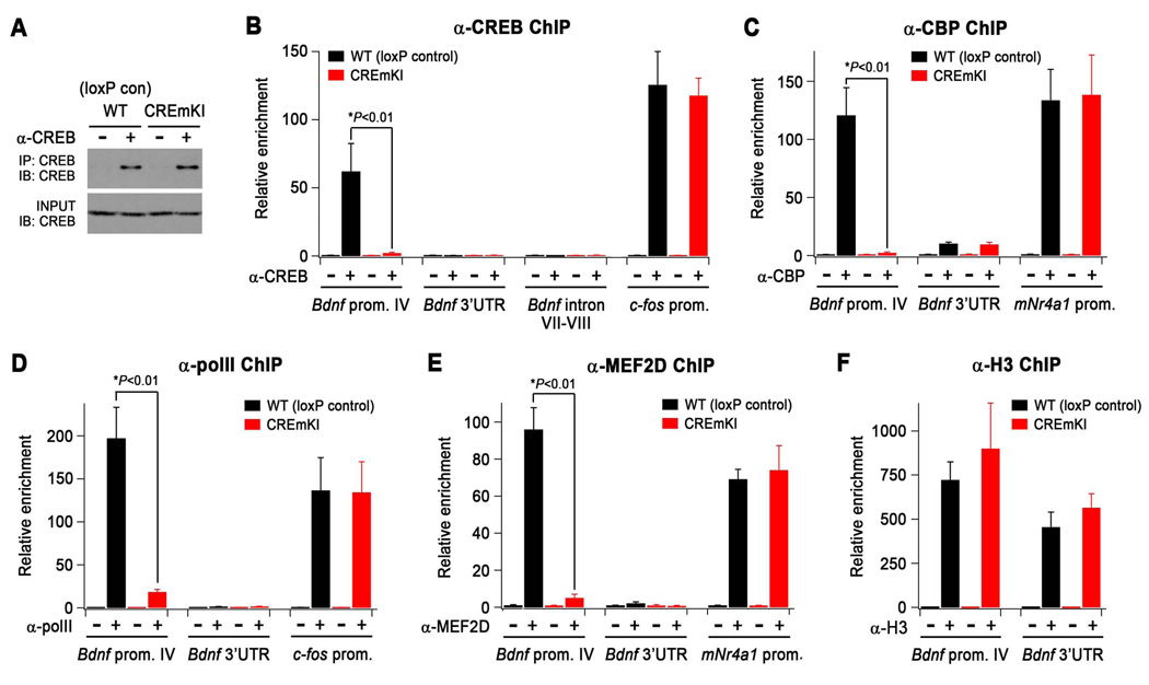
Neuronal activity-regulated gene expression has been suggested to be an important mediator of long-lasting, experience-dependent changes in the nervous system, but the activity-dependent component of gene transcription has never been selectively isolated and tested for its functional significance. Here, we demonstrate that introduction of a subtle knockin mutation into the mouse Bdnf gene that blocks the ability of the activity-regulated factor CREB to bind Bdnf promoter IV results in an animal in which the sensory experience-dependent induction of Bdnf expression is disrupted in the cortex. Neurons from these animals form fewer inhibitory synapses, have fewer spontaneous inhibitory quantal events, and exhibit reduced expression of inhibitory presynaptic markers in the cortex. These results indicate a specific requirement for activity-dependent Bdnf expression in the development of inhibition in the cortex and demonstrate that the activation of gene expression in response to experience-driven neuronal activity has important biological consequences in the nervous system.


Figures

Figure 1
Figure 1
Generation of Bdnf pIV, TMKI, CREmKI, and loxP control mice. A) Genomic structure of the rodent Bdnf gene adopted from Aid et al., 2007. The discovery of additional exons in the rodent Bdnf gene has resulted in changes in exon nomenclature; the four major transcripts now known as I, II, IV, and VI were initially described as I, II, III, and IV (in red), respectively, in Timmusk et al., 1993. B) Targeting strategy for the introduction of pIV, TMKI, CREmKI, and loxP control mutations into Bdnf promoter IV by homologous recombination. P, PciI; A, AgeI; S, SphI; 5’, 3’ Southern probes; NEO/ZEO, neomycin-zeomycin positive selection cassette; DTA, diphtheria toxin negative selection cassette. C) Southern blot analysis of PciI-digested genomic DNA from targeted ES cells using the 5’ probe indicates correct targeting of each mutation. pIV: WT, 11.6 kb; mut., 5.3 kb; TMKI, CREmKI, loxP control: WT, 11.6 kb; mut., 5.7 kb. D) Direct sequencing of PCR products amplified from genomic DNA isolated from TMKI, CREmKI, or loxP control homozygous mice using primers spanning Bdnf promoter IV.
Figure 2
Figure 2
Activity-dependent Bdnf transcription in pIV−/−, TMKI, and CREmKI cortical neurons is impaired in response to membrane depolarization. Levels of Bdnf exon IV (A–C), exon IX (coding exon) (D–F), and c-fos (G–I) mRNA in 5DIV cortical neurons prepared from pIV−/− and wildtype littermates (A,D,G), homozygous TMKI and loxP control littermates (B,E,H), or homozygous CREmKI and loxP control littermates (C,F,I) that were either left untreated or membrane depolarized with high extracellular potassium for the indicated amounts of time. Differences in Bdnf exon IV and total Bdnf mRNA expression are statistically significant for pIV−/−, TMKI, or CREmKI versus their respective controls (P<0.01, repeated-measures ANOVA; P<0.01, pairwise comparisons at each timepoint 1 hr or longer, Bonferroni-Dunn post-hoc test). Data are mean ± SEM from n=3 (pIV−/− and CREmKI) or n=2 (TMKI) independent experiments in which each sample was measured in triplicate.
Figure 3
Figure 3
Neuronal activity-dependent Bdnf transcription in CREmKI cortical neurons is impaired in response to glutamate receptor activation. Levels of Bdnf exon IV (A), total Bdnf (exon IX) (B), Bdnf exon I (C), Bdnf exon II (D), NP2 (E), and Bdnf exon VI (F) mRNA in 12DIV cortical neurons prepared from CREmKI or loxP control littermates that were either mock-stimulated or treated with 20µM NMDA for the indicated amounts of time. Asterisk denotes P<0.01, repeated-measures ANOVA, pairwise comparisons at indicated timepoints, Bonferroni-Dunn post-hoc correction. Data are mean ± SEM from n=2 independent experiments in which each sample was measured in triplicate.
Figure 4
Figure 4
Sensory experience-dependent Bdnf expression is impaired in the intact CREmKI brain. A–B) Levels of the indicated transcripts in the primary visual cortex of adult CREmKI and loxP control mice reared in darkness for 14 days, after which they were either maintained in darkness (−) or exposed to light for 90 min. (+). Within each transcript, mRNA levels are reported as fold induction relative to the loxP control unstimulated (−) condition. In either loxP control or CREmKI visual cortex, light stimulation induced the expression of all mRNAs measured (P<0.01, two-way ANOVA with Bonferroni Dunn post-hoc test), except for gapdh, which served as a non-activity-regulated gene control. C–E) Bdnf exon IV, total Bdnf, and c-fos mRNA levels in the cortex of CREmKI and loxP control mice injected with either saline (time 0) or kainic acid (KA) for the indicated amounts of time. F) Levels of BDNF protein in the primary visual cortex of animals from A–B. G) The unstimulated (−) levels of Bdnf exon IV and total Bdnf in CREmKI and loxP control visual cortex are re-plotted from A) for direct comparison (P>0.05, two-way ANOVA with Bonferroni-Dunn post-hoc test). Data are from n=5–7 animals per condition for both visual stimulation and KA seizure experiments. Each animal was measured in triplicate, and data are presented as mean ± SEM. Asterisks denote P<0.01, two-way (visual simulation) or repeated-measures (KA seizure) ANOVA with Bonferroni-Dunn post-hoc test between indicated pair.
Figure 5
Figure 5
Impaired activity-dependent Bdnf expression reduces the number of inhibitory synapses formed on CREmKI neurons in culture. A) Representative images of GFP-transfected, E17.5 + 18DIV CREmKI and loxP control littermate cortical neurons, immunostained with the indicated antibodies to label pre- and post-synaptic inhibitory synapse terminals. Scale bar, 10µm. B) Enlargement of the boxed area in A shows details of dendrites. The GFP signal is converted to gray to allow better visualization of the synaptic puncta. Scale bar, 5µm. C–E) Quantification of the average density of GAD65/GABAARγ2 (C, 78–81 cells/condition), VGAT/GABAARβ2/3 (D, 65–66 cells/condition), and synapsinI/PSD-95 (E, 63–65 cells/condition) co-clusters along the dendrites of GFP-transfected CREmKI and loxP control neurons. Asterisk denotes P<0.01, two-way ANOVA with pairwise comparison by Bonferroni-Dunn post-hoc test. Data are mean±SEM from 3–4 independent experiments. F) Quantification of dendritic branch complexity by Sholl analysis plots the number of dendritic branches intersecting concentric circles of increasing radii centered on the cell body. P>0.05 by repeated-measures ANOVA; data are mean±SEM from 26–28 cells/condition from 2 independent experiments.
Figure 6
Figure 6
Neuronal activity-dependent Bdnf expression controls the development of cortical inhibition. A) Representative traces of mIPSCs recorded from layer II/III V1 pyramidal neurons in loxP control and CREmKI acute cortical slices. B) Cumulative probability distributions of mIPSC interevent intervals (I) and amplitudes (II) recorded from loxP control and CREmKI neurons. P<0.01 by either Kolmogorov-Smirnov test or Monte Carlo simulation (see Supplemental Methods). C) Average interevent interval (I) and amplitude (II) of mIPSCs recorded from loxP control and CREmKI neurons. Data are mean ± SEM, P<0.01 by Student’s t-test. Data are from 18–20 cells/genotype recorded from 12 pairs of littermates.
Figure 7
Figure 7
Reduced immunoreactivity for inhibitory synaptic markers in CREmKI cortex. A) Representative images of immunostaining with antibodies recognizing GAD65 (red), which is enriched in inhibitory presynaptic terminals, and VGLUT1 (green), which is enriched at excitatory presynaptic terminals, in layer II/III of CREmKI and loxP control primary visual cortex. Scale bar, 5µm. B–D) Quantification of the average intensity of α-GAD65 (B, 74–77 fields/genotype), α-VGAT (C, 48 fields/genotype), and α-VGLUT1 (D, 48 fields/genotype) immunostaining in CREmKI and loxP control littermates. Asterisk indicates P<0.01, two-way ANOVA with pairwise comparison by Bonferroni-Dunn post-hoc test. Data are mean ± SEM from 3–4 pairs of littermates. E–F) Quantification of the number of paralbumin- (E) and NPY- (F) positive inhibitory neurons in the primary visual cortex of CREmKI and loxP control littermates, normalized to the number of total nuclei. P>0.05 for either parvalbumin-positive or NPY-positive neurons, two-way ANOVA. Data are mean ± SEM collected from n=24 hemispheres from 3 pairs of littermates.
Figure 8
Figure 8
Loss of CREB binding at Bdnf promoter IV in CREmKI brains leads to the disassembly of the transcriptional activating complex at Bdnf promoter IV. A) α-CREB immunoprecipitates were isolated from the cross-linked forebrains of CREmKI and loxP control littermates under conditions identical to those used for chromatin immunoprecipitation (ChIP) and analyzed by Western blotting with an independent antibody against CREB. B–F) qPCR measurement of the levels of the indicated genomic DNA regions in α-CREB (B), α-CBP(C), α-polymerase II (D), α-MEF2D (E), and α-histone H3 (F) chromatin immunoprecipitates prepared from the forebrains of CREmKI and loxP control littermates. Asterisk denotes P<0.01, two-way ANOVA with pairwise comparison by Bonferroni-Dunn post-hoc test. Data are mean ± SEM from n=4 (α-CREB), n=3 (α-MEF2D, α-CBP), or n=2 (α-pol II, α-H3) independent experiments in which each sample was measured in triplicate.

Comment in

References

    1. Abidin I, Eysel UT, Lessmann V, Mittmann T. Impaired GABAergic inhibition in the visual cortex of brain-derived neurotrophic factor heterozygous knockout mice. J Physiol. 2008;586:1885–1901. - PMC - PubMed
    1. Abidin I, Kohler T, Weiler E, Zoidl G, Eysel UT, Lessmann V, Mittmann T. Reduced presynaptic efficiency of excitatory synaptic transmission impairs LTP in the visual cortex of BDNF-heterozygous mice. Eur J Neurosci. 2006;24:3519–3531. - PubMed
    1. Aid T, Kazantseva A, Piirsoo M, Palm K, Timmusk T. Mouse and rat BDNF gene structure and expression revisited. J Neurosci Res. 2007;85:525–535. - PMC - PubMed
    1. Balschun D, Wolfer DP, Gass P, Mantamadiotis T, Welzl H, Schutz G, Frey JU, Lipp HP. Does cAMP response element-binding protein have a pivotal role in hippocampal synaptic plasticity and hippocampus-dependent memory? J Neurosci. 2003;23:6304–6314. - PMC - PubMed
    1. Bartel DP, Sheng M, Lau LF, Greenberg ME. Growth factors and membrane depolarization activate distinct programs of early response gene expression: dissociation of fos and jun induction. Genes Dev. 1989;3:304–313. - PubMed

Publication types

MeSH terms

Substances