. 2008 Nov 26;60(4):672-82.
doi: 10.1016/j.neuron.2008.10.042.

PDF cells are a GABA-responsive wake-promoting component of the Drosophila sleep circuit

Affiliations

PDF cells are a GABA-responsive wake-promoting component of the Drosophila sleep circuit

Katherine M Parisky et al. Neuron. .

Erratum in

  • Neuron. 2009 Jan 15;61(1):152. Kang, Keongjin [corrected to Kang, Kyeongjin]

Abstract

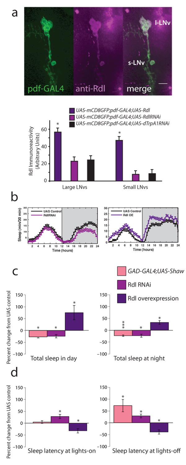
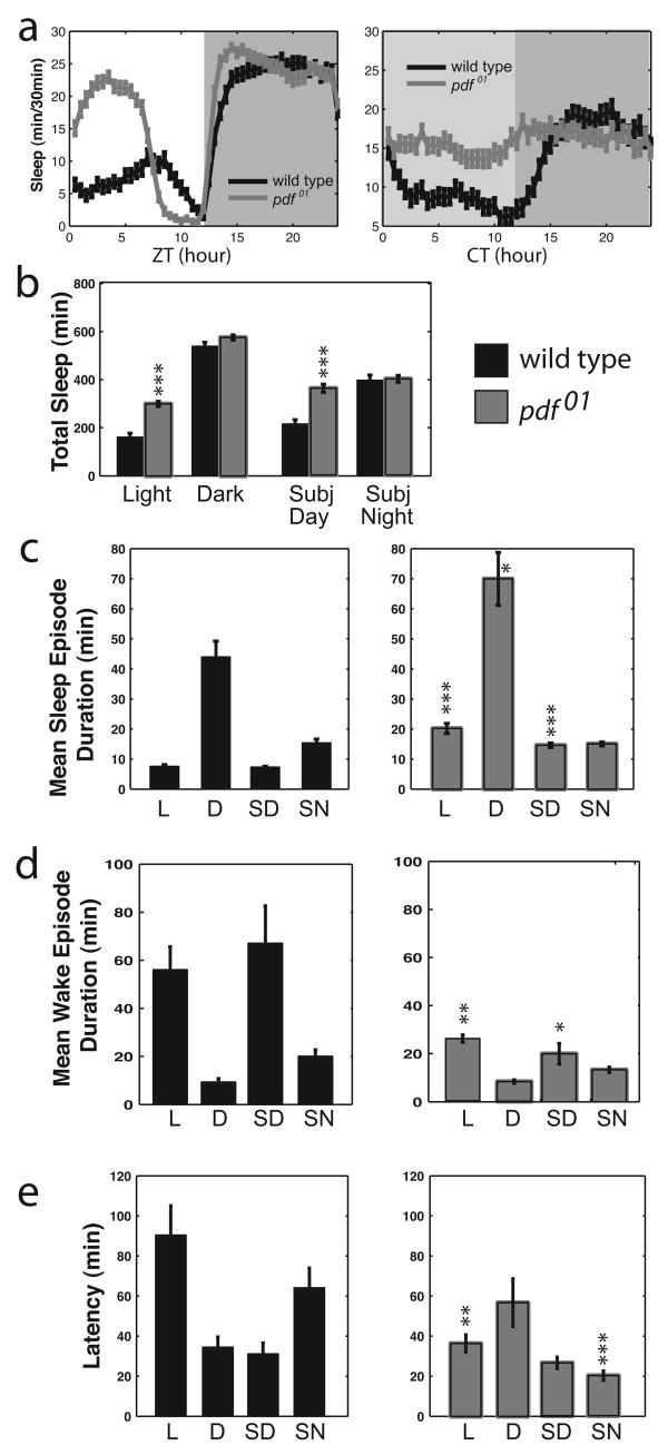
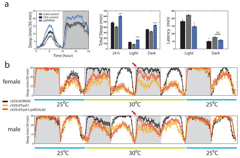
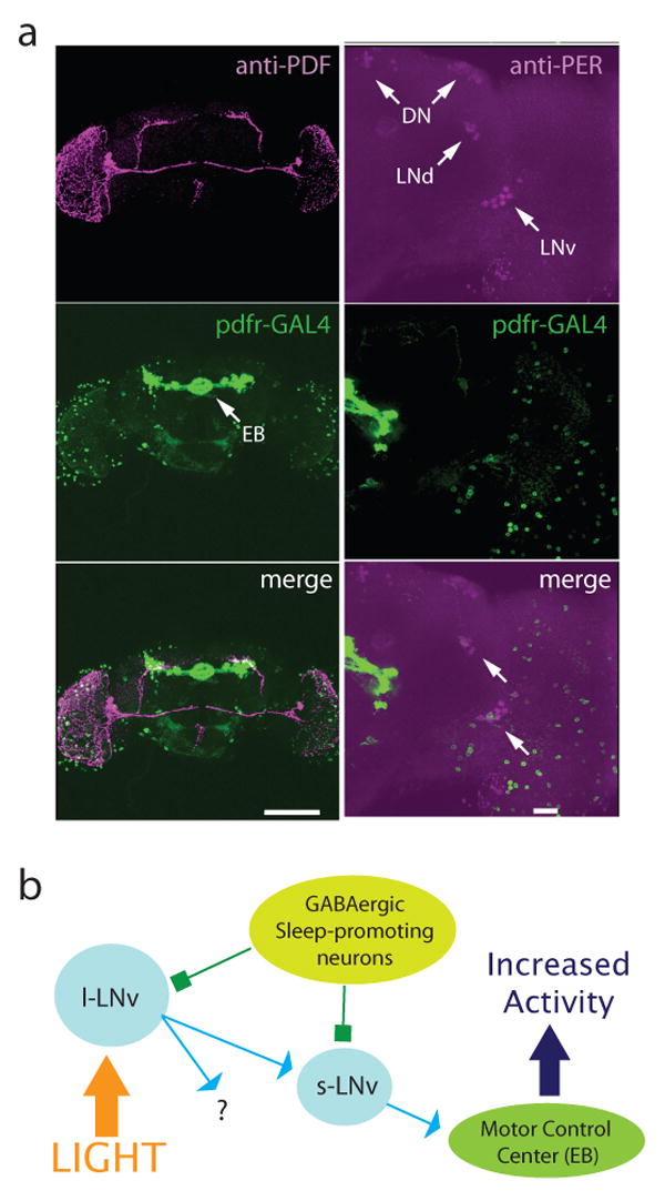
Daily sleep cycles in humans are driven by a complex circuit within which GABAergic sleep-promoting neurons oppose arousal. Drosophila sleep has recently been shown to be controlled by GABA, which acts on unknown cells expressing the Rdl GABAA receptor. We identify here the relevant Rdl-containing cells as PDF-expressing small and large ventral lateral neurons (LNvs) of the circadian clock. LNv activity regulates total sleep as well as the rate of sleep onset; both large and small LNvs are part of the sleep circuit. Flies mutant for pdf or its receptor are hypersomnolent, and PDF acts on the LNvs themselves to control sleep. These features of the Drosophila sleep circuit, GABAergic control of onset and maintenance as well as peptidergic control of arousal, support the idea that features of sleep-circuit architecture as well as the mechanisms governing the behavioral transitions between sleep and wake are conserved between mammals and insects.


Figures

Figure 1
Figure 1
LNvs express the Rdl GABAA receptor and mediate GABAergic effects on sleep. a, Top shows images of Rdl expression in wild type LNvs. Adult brains from pdf-GAL4;UAS-mCD8-GFP animals were stained with anti-Rdl (1:100) and visualized with confocal microscopy. Rdl is shown in magenta, GFP in green, overlap in white. Scale bar = 10 μm. Bottom shows quantification of somatic Rdl levels in LNvs expressing excess Rdl, RdlRNAi or control dTrpA1RNAi. b, Standard sleep plots of control and experimental flies in 12 hour: 12 hour light:dark (LD). Left panel shows the effects of reducing Rdl levels in LNvs: pdf-GAL4;UAS-RdlRNAi, right panel shows the effects of overexpressing Rdl in LNvs: pdf-GAL4;UAS-Rdl. c, GABA regulates total sleep. 12 h sleep from the light (left) or dark (right) period in LD was assessed for animals with decreased overall GABAergic transmission (GAD-GAL4;UAS-Shaw; n=62), decreased LNv Rdl levels (pdf-GAL4;UAS-RdlRNAi; n = 93), or increased LNv Rdl levels (pdf-GAL4;UAS-Rdl; n = 21). Data are expressed as the percent change from the genetic control. d, GABA regulates sleep onset. The latency to first sleep bout during the light (left) or dark (right) period in LD was assessed for the same genotypes. Data are expressed as the percent change from the genetic control. * indicates P < 0.05, ** P < 0.005 and *** P < 0.0005 for comparisons of experimental and control using ANOVA with Tukey posthoc test.
Figure 2
Figure 2
Shaw RNAi and dominant negative Na+/K+-ATPase increase neuronal excitability. a, Shaw RNAi reduces endogenous Shaw expression. (left) Expression of Shaw double-stranded RNA in adult central and motor neurons (GAL4-C380/UAS-ShawRNAi) causes reduction in Shaw levels. Endogenous Shaw levels were detected with the anti-C terminus Shaw antibody and quantified in the mushroom body calyx region with Leica confocal software. A significant reduction (P < 0.05) in intensity (arbitrary units) is seen in central neurons expressing ShawRNAi. (right) Expression of interfering Shaw double-stranded RNA (GAL4-24B/UAS-ShawRNAi) decreases endogenous Shaw compared to controls (+/UAS-ShawRNAi) when compared by immunoblot. Whole head lysates were separated by SDS-PAGE, transferred to nitrocellulose membranes and detected with a antibody (1:1000) to the C-terminus of Shaw which detects full-length Shaw protein (Hodge et al., 2005). Anti-Tubulin (1:200,000) was used to assess protein loading. b, Expression of Shaw RNAi in larval motor neurons with C380-GAL4 increases excitability. (left) Traces of whole cell current clamp recording from MNISN-Is of control (C380-GAL4 only) and experimental (C380-GAL4;UAS-ShawRNAi) animals injected with 60 pA current. (right) Quantified data for the firing rate response to various current injections. n = 7 for C380-GAL4 alone and n = 10 for C380-GAL4;UAS-ShawRNAi. c, Expression of dominant negative Na+/K+-ATPase in larval motor neurons with C380-GAL4 increases excitability. (left) Traces of whole cell current clamp recording from MNISN-Is of control (C380-GAL4 only) and experimental (C380-GAL4;UAS-dnATPase) animals injected with 60 pA current. (right) Quantified data for the firing rate response to various current injections. n = 8 for C380-GAL4 alone and n = 7 for C380-GAL4;UAS-dnATPase.
Figure 3
Figure 3
Excitability of LNvs controls sleep. a, Standard sleep plots of control and experimental flies in 12 hour: 12 hour light:dark (LD) or constant darkness (DD). Top panel show the effects of reducing neuronal activity levels in LNvs: pdf-GAL4;UAS-EKO. Bottom panels show the effects of enhancing normally patterned activity in LNvs: pdf-GAL4;UAS-dnATPase. b, LNv activity controls total sleep. 12 h sleep from the light (left) or dark (right) period in LD was assessed for animals with suppressed responsiveness to inputs (pdf-GAL4;UAS-EKO; n = 55), or increased responsiveness to inputs (pdf-GAL4;UAS-ShawRNAi and pdf-GAL4;UAS-dnATPase; n = 32 and 80). Data are expressed as the percent change from the genetic control. c, LNv activity controls sleep onset. The latency to first sleep bout during the light (left) or dark (right) period in LD was assessed for the same genotypes. Data are expressed as the percent change from the genetic control. d, LNvs mediate the wake-promoting effects of light. Latency to first sleep bout during the light period in LD (L) or subjective day in DD (SD) is shown for animals with reduced responsiveness to inputs (left, pdf-GAL4;UAS-EKO) or with increased responsiveness to inputs (right, pdf-GAL4;UAS-dnATPase). e, Acute activation of LNvs disrupts nighttime sleep. pdf-GAL4;UAS-dTrpA1 and control animals (n = 32 for each genotype) were raised at the non-permissive temperature of 22°C and entrained in LD at that temperature. Data were collected for 3 days then temperature was increased to 27°C to activate dTrpA1. Left panel shows the 3 days immediately preceding the temperature increase. Middle panel shows 3 days after temperature increase. Left panel shows arousal state stability at 27°C for all genotypes in the early evening (ZT12-15; time marked by arrow in middle panel). * indicates P < 0.05, ** P < 0.005 and *** P < 0.0005 for t-test comparisons of experimental and control in panels b and c and for Tukey post-hoc test after ANOVA for panels d and e.
Figure 4
Figure 4
pdf01 mutants have increased total sleep and decreased sleep latency. a, Standard sleep plots of control and mutant flies in 12 hour: 12 hour light:dark (LD, left) or in constant darkness (DD, right). b, Total sleep for controls (black bars) and pdf01 mutants (gray bars) for the light period and dark period in LD and subjective day and subjective night in DD. c, Mean sleep episode duration, d, Mean wake episode duration, and e, Latency to first sleep bout, for control (left) and pdf01 mutants (right). Data are shown for light (L) and dark (D) periods in LD and for subjective day (SD) and subjective night (SN) in DD. Data are presented as means ± SEM. * indicates P < 0.05, ** P < 0.005, *** P < 0.0005 for the comparison to control by ANOVA with Tukey posthoc test. n = 106.
Figure 5
Figure 5
Both large and small LNvs are involved in sleep control. a, Down regulation of the PDFR with UAS-pdfrRNAi driven by pdf-GAL4 in LNvs increases both daytime and nighttime sleep, but only significantly affects daytime latency. Standard sleep plots of female flies in 12 hour: 12 hour light:dark are shown. * indicates P < 0.05, *** indicates P < 0.0005 and ns indicates “not significant” for the comparison to other genotypes by ANOVA with Tukey posthoc test. n = 70, 71 and 75 for UAS alone (UAS-pdfrRNAi), GAL4 alone (pdfGAL4) and experimental (pdf-GAL4;UAS-pdfrRNAi) respectively. b, Continuous sleep data from flies expressing the temperature-gated cation channel dTrpA1 in peptidergic neurons ± l-LNvs. Flies were entrained in LD for 5 days at 25°C (last day is shown) and shifted to 30°C for two days, then back to 25°C. Females (n = 16 for control c929-GAL4;UAS-dORKNC), 14 for c929-GAL4;UAS-dTrpA1 and 21 for c929-GAL4;pdf-GAL80;UAS-dTrpA1) are shown at top, males (n = 23 for control, 20 for c929 and 19 for c929+pdf-GAL80) at bottom. Arrow indicates rescue of early evening sleep by suppression of dTrpA1 expression in l-LNvs by pdf-GAL80 on day 2 of elevated temperature.
Figure 6
Figure 6
Output of the sleep circuit. a, pdfr-GAL4 marks output cells of the LNv circuit. Left panels show reconstructed pdfr-GAL4;UAS-mCD8-GFP adult brain stained with anti-PDF (1:1000) and visualized with confocal microscopy. PDF is shown in magenta, GFP in green, overlap in white. Scale bar = 150 μm. Right panels show a 37 μ section of a pdfr-GAL4;UAS-mCD8-GFP adult brain stained with anti-Per (1:100) and visualized with confocal microscopy. Per is shown in magenta, GFP in green, overlap in white. Arrows indicate clock cells. Scale bar = 20 μm. b, Model of the Drosophila sleep circuit. Light, and perhaps other arousal cues, activate l-LNvs which release PDF onto s-LNvs that project to other clock cells and also send dorsal projections that pass by pdfr-GAL4 positive cells groups such as the ellipsoid bodies that are involved in control of activity. Both l- and s-LNvs express GABAA receptors, allowing sleep promoting GABAergic neurons to suppress wakefulness.

References

    1. Agosto J, Choi JC, Parisky KM, Stilwell G, Rosbash M, Griffith LC. Modulation of GABAA receptor desensitization uncouples sleep onset and maintenance in Drosophila. Nat Neurosci. 2008;11:354–359. - PMC - PubMed
    1. Allada R, White NE, So WV, Hall JC, Rosbash M. A mutant Drosophila homolog of mammalian Clock disrupts circadian rhythms and transcription of period and timeless. Cell. 1998;93:791–804. - PubMed
    1. Borbely AA, Achermann P. Sleep homeostasis and models of sleep regulation. J Biol Rhythms. 1999;14:557–568. - PubMed
    1. Broughton SJ, Kitamoto T, Greenspan RJ. Excitatory and inhibitory switches for courtship in the brain of Drosophila melanogaster. Curr Biol. 2004;14:538–547. - PubMed
    1. Choi JC, Park D, Griffith LC. Electrophysiological and morphological characterization of identified motor neurons in the Drosophila third instar larva central nervous system. J Neurophysiol. 2004;91:2353–2365. - PubMed

Publication types

MeSH terms