Review
. 2009 Feb 15;315(4):638-48.
doi: 10.1016/j.yexcr.2008.10.024. Epub 2008 Oct 31.

Ligand-induced ErbB receptor dimerization

Affiliations
Review

Ligand-induced ErbB receptor dimerization

Mark A Lemmon. Exp Cell Res. .

Abstract

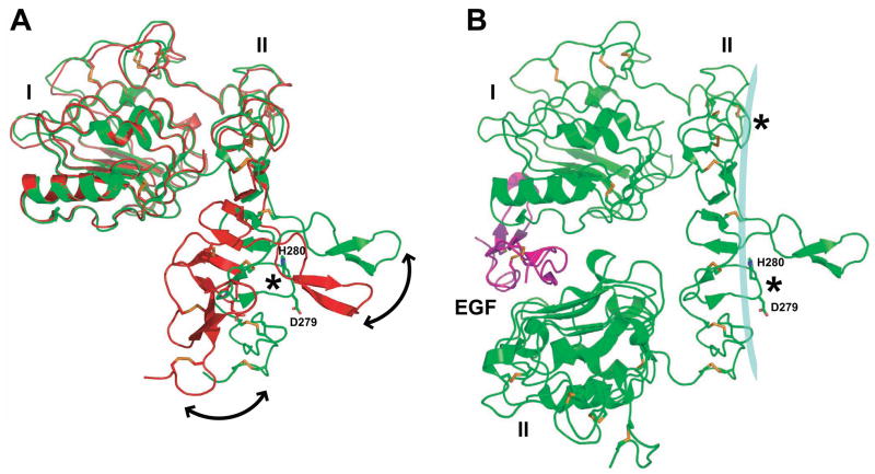
Structural studies have provided important new insights into how ligand binding promotes homodimerization and activation of the EGF receptor and the other members of the ErbB family of receptor tyrosine kinases. These structures have also suggested possible explanations for the unique properties of ErbB2, which has no known ligand and can cause cell transformation (and tumorigenesis) by simple overexpression. In parallel with these advances, studies of the EGF receptor at the cell surface increasingly argue that the structural studies are missing key mechanistic components. This is particularly evident in the structural prediction that EGF binding linked to receptor dimerization should be positively cooperative, whereas cell-surface EGF-binding studies suggest negative cooperativity. In this review, I summarize studies of ErbB receptor extracellular regions in solution and of intact receptors at the cell surface, and attempt to reconcile the differences suggested by the two approaches. By combining results obtained with receptor 'parts', it is qualitatively possible to explain some models for the properties of the whole receptor. These considerations underline the need to consider the intact ErbB receptors as intact allosterically regulated enzymes, and to combine cellular and structural studies into a complete picture.


Figures

Figure 1
Figure 1
Model for EGF-induced dimerization of the EGFR extracellular region. The top panel shows ribbon representations of sEGFR structures with- and without bound EGF. The left-hand structure is from Ferguson et al. [14], and shows the domain II/IV tether (ringed with orange oval) that occludes the dimerization arm. EGF binding to this structure induces a conformational change that can be modeled approximately by a 130° rotation of the domain I/II fragment about the axis between domains II and III marked by the black circle, in addition to a 20Å translation into the page. This change causes EGFR to adopt the extended conformation, in which the dimerization arm is exposed to drive dimerization as shown in the right-hand panel. The sEGFR dimer structure is based on the coordinates of the EGF/sEGFR complex published by Ogiso et al. [20]. We have modeled domain IV in this structure based on the domain III/IV relationship seen in unliganded sEGFR. Dimerization arm contacts at the dimer interface are ringed with an orange oval. Domains I and III are colored red and red/grey respectively. Domains II and IV are colored green and green/grey respectively. EGF is cyan. The lower panel shows a cartoon representation of this dimerization reaction, with each domain presented as a red or green rectangle.
Figure 2
Figure 2
Structures of all four human ErbB receptor extracellular regions without bound ligand. EGFR, ErbB3 and ErbB4 all adopt the tethered conformation in the absence of ligand, whereas ErbB2 adopts a tethered conformation that resembles the ligand-activated, dimerization-competent, EGFR protomers in the EGFR dimer shown in Figure 1. Structures are shown in ribbon representation, with colors as described for Figure 1. The sEGFR structure is from Li et al. [53], sErbB2 is from Cho et al. [15], sErbB3 from Cho and Leahy [18], and sErbB4 from Bouyain et al. [21].
Figure 3
Figure 3
Remodeling of the domain II dimerization interface of EGFR upon EGF binding. In panel A, the domain I/II fragments of sEGFR without bound ligand (1YY9 [53], shown in red) and with bound EGF (1IVO [20], shown in green) are overlaid with domain I as the reference. Although the first four disulfide-bonded modules of domain II overlay very well between the two structures, the trajectory of the domain is altered substantially beyond a point immediately N-terminal to the dimerization arm. As a result, the dimerization arm is reoriented substantially as shown by a double-headed black arrow. The C-terminus of domain II (where it links to domain III) is also repositioned significantly (also marked by double-headed curved arrow). As a result, the dimer contact site involving D279 and H280 (marked with an asterisk) lies in a very different position in the red inactive structure than in the green active structure. The different curvature imposed on domain II is thus proposed to ‘remodel’ the dimerization interface so that multiple weak contacts can cooperate with the dimerization arm in driving high affinity dimerization as described in the text. In B, one monomer from the EGF-bound sEGFR dimer [20] is shown. As mentioned in the legend to Figure 1, much of domain IV was missing from this structure. EGF is magenta, sEGFR is green. D279 and H280, which make crucial contacts across the dimerization interface [52], are marked. As discussed in the text, EGF (or another ligand) is thought to impose a precise bend on domain II (shown as a cyan curve against domain II) that defines the nature of the dimerization interface. Different ligands might promote slightly different domain I/III relationships, and thus impose subtly different bends on domain II.
Figure 4
Figure 4
Equilibrium scheme for EGF binding to EGFR and EGFR dimerization. For ligand binding, K1 refers to EGF association with monomeric EGFR (to form an RL complex, where R is Receptor and L is Ligand). K2 refers to EGF binding to an already-dimerized EGFR (R2) to form a singly-occupied R2L dimer. K3 refers to EGF binding to the R2L complex to yield a fully occupied R2L2 dimer. Kα denotes the association constant for free EGFR (to generate R2). Kβ describes EGFR association with a monomeric EGF:EGFR complex to yield the R2L complex. Kγ reflects dimerization of the EGF:EGFR complex to form an R2L2 dimer. The system can be described completely with any four of the listed association constants. According to Macdonald and Pike [75] and Wofsy et al. [67], the concave-up Scatchard plots seen when EGF binding to cell-surface EGFR is analyzed can be explained if K2>K3i.e. if EGF binds more strongly to an R2 dimer than to an R2L dimer, so that the second ligand binding event is weaker than the first. If K2 = K3, the model predicts positive cooperativity [–69, 75]. In addition, K2 must be greater than K1 in order for EGF binding to drive dimerization. In models that predict negative cooperativity, the R2L species must accumulate, as discussed in the text. Loss of domains that stabilize the R2L species may explain the failure to see negative cooperativity with the isolated EGFR extracellular region. In the model of Macdonald and Pike [75], values of Kα and Kβ are similar, providing a potential explanation for the significant ligand-independent EGFR dimerization described in the text. In this model, Kγ is reduced by ~10-fold, suggesting that binding of a second ligand molecule to the R2L species reduces the stability of the dimer. Rather than imposing negative cooperativity, Klein et al. [69] assumed in their analysis that the binding events represented by K2 and K3 were equivalent – citing the symmetric nature of the EGF:sEGFR dimeric complex. With this model, the experimentally observed concave-up Scatchard curvature can be modeled by including an ‘external site’ that stabilizes a subset of occupied EGFR dimers.

References

    1. Ullrich A, Schlessinger J. Signal transduction by receptors with tyrosine kinase activity. Cell. 1990;61:203–212. - PubMed
    1. Yarden Y, Schlessinger J. Epidermal growth factor induces rapid, reversible aggregation of the purified epidermal growth factor receptor. Biochemistry. 1987;26:1443–1451. - PubMed
    1. Yarden Y, Schlessinger J. Self-phosphorylation of epidermal growth factor receptor: evidence for a model of intermolecular allosteric activation. Biochemistry. 1987;26:1434–1442. - PubMed
    1. Jorissen RN, Walker F, Pouliot N, Garrett TPJ, Ward CW, Burgess AW. Epidermal growth factor receptor: mechanisms of activation and signalling. Exp Cell Res. 2003;284:31–53. - PubMed
    1. Holbro T, Hynes NE. ErbB receptors: directing key signaling networks throughout life. Annu Rev Pharmacol Toxicol. 2004;44:195–217. - PubMed

Publication types