. 2009 Feb 15;44(4):1324-33.
doi: 10.1016/j.neuroimage.2008.10.037. Epub 2008 Nov 7.

Reliability of MRI-derived cortical and subcortical morphometric measures: effects of pulse sequence, voxel geometry, and parallel imaging

Affiliations

Reliability of MRI-derived cortical and subcortical morphometric measures: effects of pulse sequence, voxel geometry, and parallel imaging

J S Wonderlick et al. Neuroimage. .

Abstract

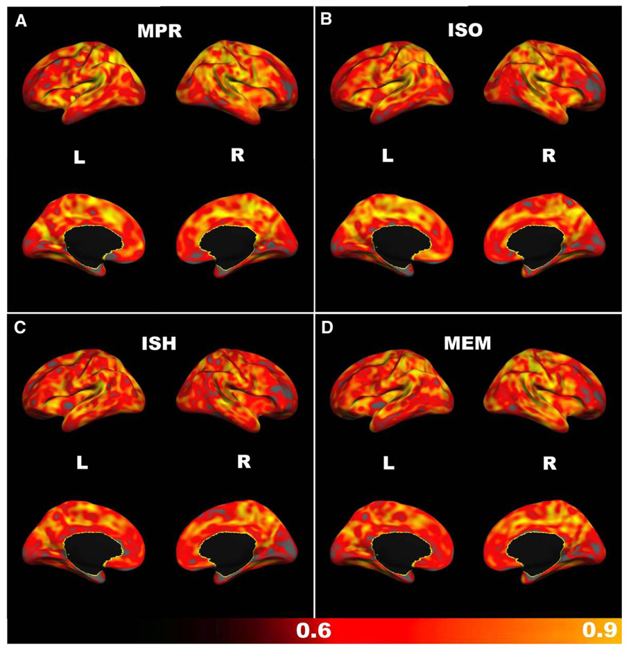
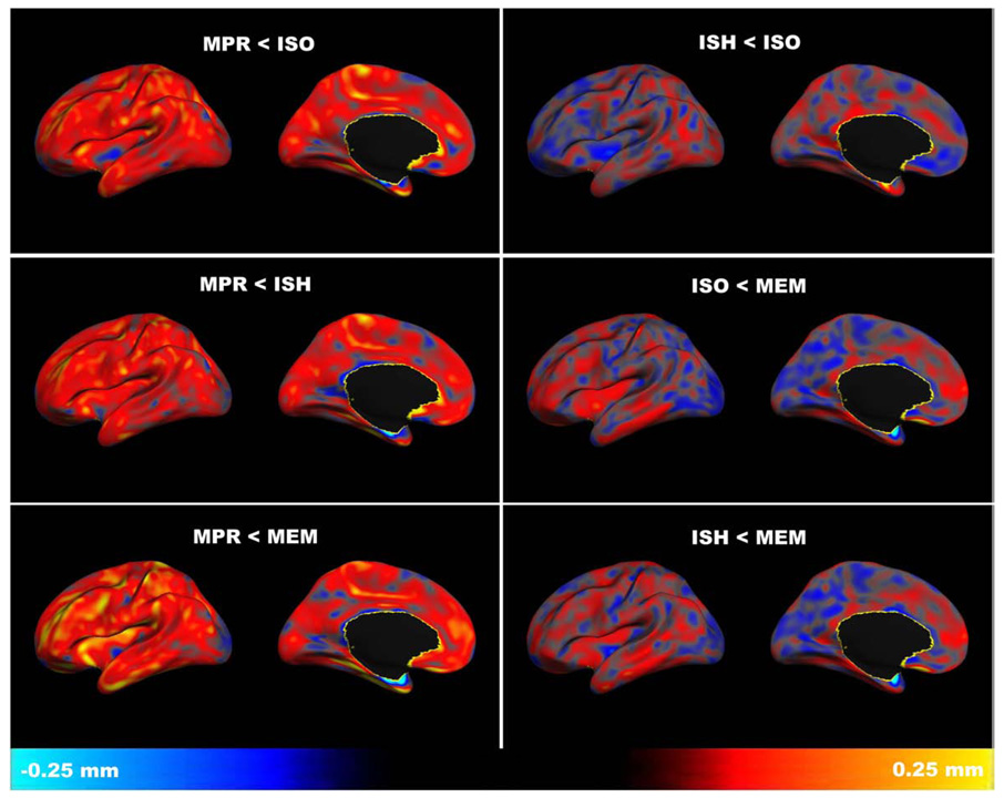
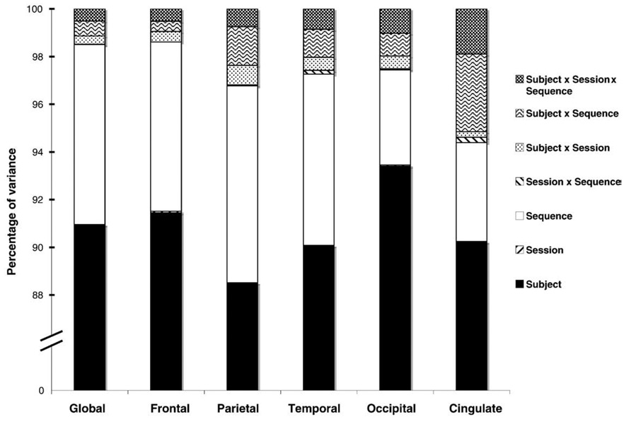
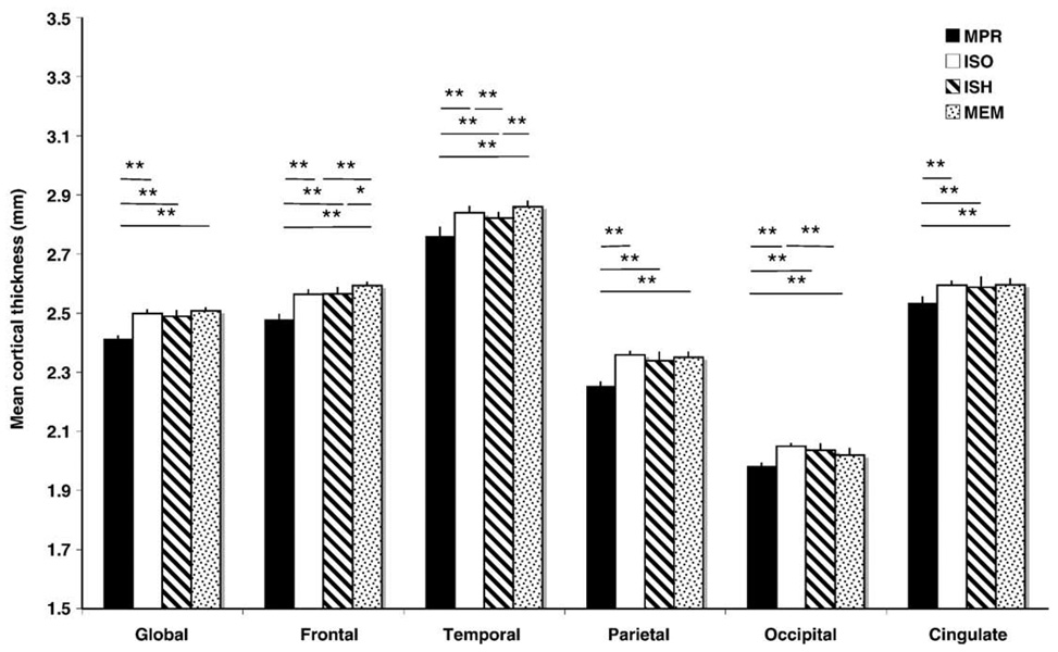
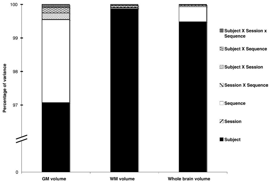
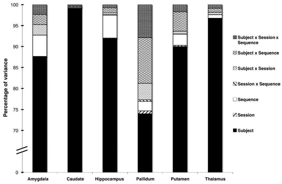
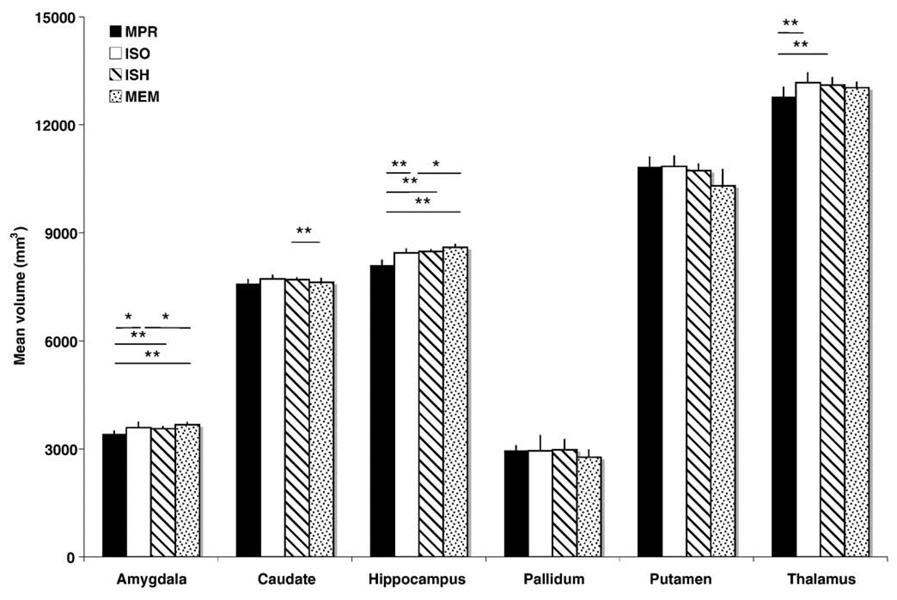
Advances in magnetic resonance imaging (MRI) have contributed greatly to the study of neurodegenerative processes, psychiatric disorders, and normal human development, but the effect of such improvements on the reliability of downstream morphometric measures has not been extensively studied. We examined how MRI-derived neurostructural measures are affected by three technological advancements: parallel acceleration, increased spatial resolution, and the use of a high bandwidth multiecho sequence. Test-retest data were collected from 11 healthy participants during 2 imaging sessions occurring approximately 2 weeks apart. We acquired 4 T1-weighted MP-RAGE sequences during each session: a non-accelerated anisotropic sequence (MPR), a non-accelerated isotropic sequence (ISO), an accelerated isotropic sequence (ISH), and an accelerated isotropic high bandwidth multiecho sequence (MEM). Cortical thickness and volumetric measures were computed for each sequence to assess test-retest reliability and measurement bias. Reliability was extremely high for most measures and similar across imaging parameters. Significant measurement bias was observed, however, between MPR and all isotropic sequences for all cortical regions and some subcortical structures. These results suggest that these improvements in MRI acquisition technology do not compromise data reproducibility, but that consistency should be maintained in choosing imaging parameters for structural MRI studies.


Conflict of interest statement

Disclosure statement

The authors have no actual or potential conflicts of interest.

Figures

Fig. 1
Fig. 1
Cortical thickness: intra-class correlation coefficients (ICCs) across scanning sessions, calculated for each vertex on the cortical surface. Gray areas represent ICC values of less than 0.6. (A) anisotropic MP-RAGE, (B) isotropic MP-RAGE, (C) accelerated MP-RAGE, (D) accelerated isotropic multiecho MP-RAGE.
Fig. 2
Fig. 2
Cortical thickness: mean difference between sequences, calculated for each vertex on the cortical surface (left hemisphere shown; right hemisphere is similar). Comparisons between anisotropic and isotropic sequences reveal a bias towards smaller measurements in the anisotropic MP-RAGE (MPR) relative to the isotropic single-echo MP-RAGE (ISO), accelerated isotropic single-echo MP-RAGE (ISH), and accelerated isotropic multiecho MP-RAGE (MEM) sequences. Cortical thickness differences tend to be smaller and more evenly distributed between isotropic sequences (ISO
Fig. 3
Fig. 3
Percentage of variance for cortical thickness measures by factor and interaction. Subject and sequence factors accounted for the majority (>90%) of variance in these measures, indicating a high degree of test-retest reliability.
Fig. 4
Fig. 4
Mean cortical thickness measures by sequence (*p<0.05, **p<0.01, Bonferroni corrected). Error bars indicate mean cortical thickness difference across sessions. Measures derived from the anisotropic MP-RAGE (MPR) were significantly lower compared to all other sequences for all cortical regions. Multiecho MP-RAGE (MEM) measures tended to be higher in frontal and temporal regions, but lower in the occipital lobe. In addition, the temporal cortical thickness metric was significantly decreased for accelerated isotropic MP-RAGE (ISH) measures compared to non-accelerated isotropic MP-RAGE (ISO).
Fig. 5
Fig. 5
Percentage of variance for brain volume measures by factor and interaction. Subject and sequence factors accounted for greater than 99.5% of total variance for surface-based measures of brain volume. For measures of WM volume, where a measurement bias between sequences was not observed, subject factors alone accounted for almost all variance.
Fig. 6
Fig. 6
Percentage of variance in segmented measures of volume by structure. Subject factors contributed most to the variance (>70%) for all measures. For measures of caudate and thalamic volume, which were among the most reliable examined, variance consisted almost entirely of between-subject differences. Hippocampal and amygdalar measures were also highly reliable, with differences between sequences accounting for somewhat more variance than for the caudate or thalamus. Measures of pallidal volume were among the least reliable examined, with interactions between subject × sequence, subject × session, and subject × sequence × session contributing noticeably to overall variance.
Fig. 7
Fig. 7
Mean segmented volume of structures by sequence (*p<0.05, **p<0.01, Bonferroni corrected). Error bars indicate mean volume differences across sessions. Anisotropic MP-RAGE (MPR) measures were significantly lower for the amygdala, hippocampus, and thalamus compared to all other sequences, whereas multiecho MPRAGE (MEM) measures were significantly higher compared to the non-accelerated isotropic MP-RAGE (ISO).

References

    1. Augustinack JC, van der Kouwe AJ, Blackwell ML, Salat DH, Wiggins CJ, Frosch MP, Wiggins GC, Potthast A, Wald LL, Fischl BR. Detection of entorhinal layer II using 7 Tesla [corrected] magnetic resonance imaging. Ann. Neurol. 2005;57:489–494. - PMC - PubMed
    1. Carlson JW, Minemura T. Imaging time reduction through multiple receiver coil data acquisition and image reconstruction. Magn. Reson. Med. 1993;29:681–687. - PubMed
    1. Dale AM, Fischl B, Sereno MI. Cortical surface-based analysis. I. Segmentation and surface reconstruction. Neuroimage. 1999;9:179–194. - PubMed
    1. Deichmann R, Good CD, Josephs O, Ashburner J, Turner R. Optimization of 3-D MP-RAGE sequences for structural brain imaging. Neuroimage. 2000;12:112–127. - PubMed
    1. Desikan RS, Segonne F, Fischl B, Quinn BT, Dickerson BC, Blacker D, Buckner RL, Dale AM, Maguire RP, Hyman BT, Albert MS, Killiany RJ. An automated labeling system for subdividing the human cerebral cortex on MRI scans into gyral based regions of interest. Neuroimage. 2006;31:968–980. - PubMed

Publication types