. 2009 Feb 5;384(1):114-24.
doi: 10.1016/j.virol.2008.10.035. Epub 2008 Nov 28.

SV40 lymphomagenesis in Syrian golden hamsters

Affiliations

SV40 lymphomagenesis in Syrian golden hamsters

Adrienne L McNees et al. Virology. .

Abstract

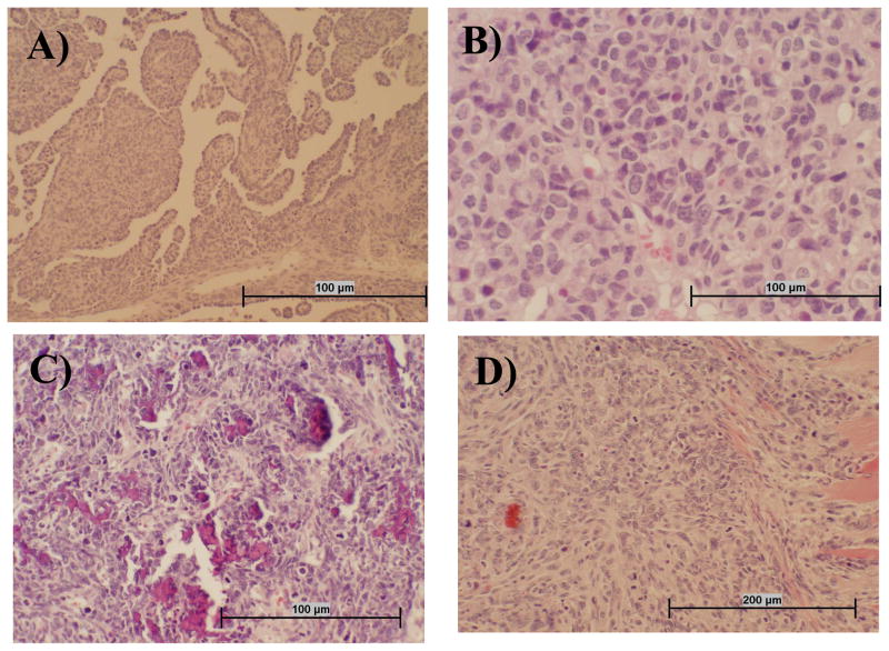
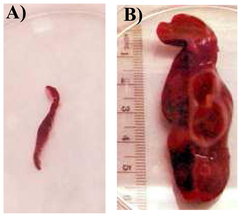
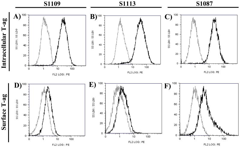
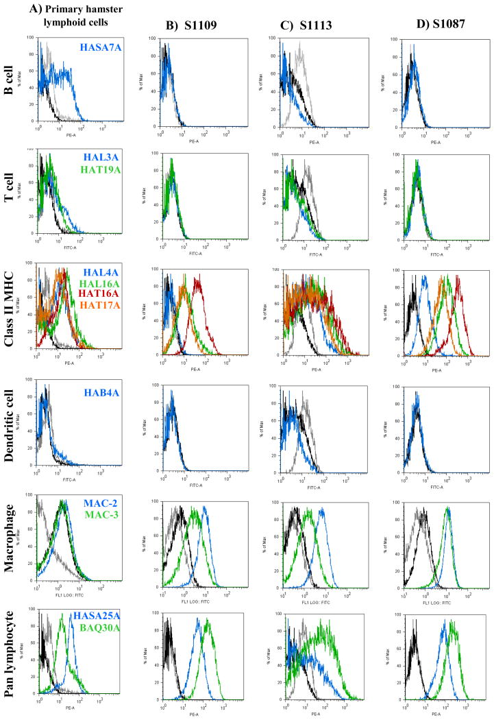
Simian virus 40 (SV40) isolates differ in oncogenic potential in Syrian golden hamsters following intraperitoneal inoculation. Here we describe the effect of intravenous exposure on tumor induction by SV40. Strains SVCPC (simple regulatory region) and VA45-54(2E) (complex regulatory region) were highly oncogenic following intravenous inoculation, producing a spectrum of tumor types. Three lymphoma cell lines were established; all expressed SV40 T-antigen, were immortalized for growth in culture, and were tumorigenic following transplantation in vivo. New monoclonal antibodies directed against hamster lymphocyte surface antigens are described. The cell lines expressed MHC class II and macrophage markers and were highly phagocytic, indicating a histiocytic origin. Many hamsters that remained tumor-free developed SV40 T-antigen antibodies, suggesting that viral replication occurred. This study shows that route of exposure influences the pathogenesis of SV40-mediated carcinogenesis, that SV40 strain VA45-54(2E) is lymphomagenic in hamsters, that hamster lymphoid cells of histiocytic origin can be transformed in vivo and established in culture, and that reagents to hamster leukocyte differentiation molecules are now available.


Figures

Fig. 1
Fig. 1
Representative tumor types that developed following i.v. inoculation of SV40. Sections of hamster tissue were collected at necropsy, formalin fixed, and stained with hematoxylin and eosin. Panel A: Mesothelioma tissue is characterized by infiltrative mass of atypical round to spindle cells often having papillary structures that are lined in one layer of round to cuboidal medium-sized cells with prominent nuclei. Panel B: Lymphoma tissue appears as highly cellular tumor tissue composed of homogeneous round cells that often have an indented nucleus and multiple nucleoli. Panel C: Osteosarcoma tissue is composed of atypical pleomorphic cells that are stellate, pyriform, and polyhedral, with tumor cells often oriented around small spicules of pale osteoid. There are also numerous fragments of bone in this section that are not decalcified. Panel D: The spindle cell carcinoma shown has an infiltrative mass into muscle composed of atypical enlongate to spindle cells with prominent nuclei and multiple nucleoli.
Fig. 2
Fig. 2
Spleens harvested from tumor-free hamsters (A) and 1113 (B), each at 8 months after i.v. inoculation with SV40 strain 776-VA(2E).
Fig. 3
Fig. 3
Expression of SV40 T-ag detected by intracellular staining or surface staining and flow cytometry on hamster lymphoma cell lines S1109 (Panels A, D), S1113 (Panels B, E), and S1087 (Panels C, F). Intracellular expression (Panels A–C) was detected using PAb101 (black lines); negative control staining was done with mouse IgG2a (gray lines). Surface expression (Panels D–F) of SV40 T-ag was detected using PAb416 (black lines); negative control staining was done with IgG2a (gray lines).
Fig. 4
Fig. 4
Surface expression of epitopes specific for lymphocyte subpopulations detected by flow cytometry. Panel A: primary hamster lymphoid cells; Panels B-D: established SV40-positive hamster lymphoma cell lines. For all histograms, negative controls are shown by gray lines representing unstained cells and black lines representing cells stained only with secondary antibody. Cells stained with the specific anti-hamster antibodies are color-coded to the histograms. Primary splenocytes from uninoculated hamsters were used as normal controls for assays with all monoclonal antibodies except MAC-2, for which primary hamster peripheral blood mononuclear cells were used as controls.
Fig. 5
Fig. 5
Phagocytosis by hamster lymphoma cell lines measured by flow cytometry. The percentage of cells that internalized fluorescent-labeled beads, averaged from two independent experiments, are graphed with standard error of the mean. Control samples were incubated at 4 °C. The average viability for each sample from both experiments is shown below the graph.
Fig. 6
Fig. 6
Internalization of phagocytosed beads by hamster lymphoma cell lines measured by confocal microscopy. Cells are shown 120 min after the addition of 30 fluorescent-labeled beads per cell and incubation at either 4°C (Panels A, C, E) or 37 °C (Panels B, D, F). Shown are hamster cell lines S1109 (Panels A, B), S1113 (Panels C, D), and S1087 (Panels E, F). Original magnification, 20×.

Similar articles

Cited by

References

    1. Amara K, Trimeche M, Ziadi S, Laatiri A, Hachana M, Sriha B, Mokni M, Korbi S. Presence of simian virus 40 DNA sequences in diffuse large B-cell lymphomas in Tunisia correlates with aberrant promoter hypermethylation of multiple tumor suppressor genes. Int J Cancer. 2007;121:2693–2702. - PubMed
    1. Arrington AS, Moore MS, Butel JS. SV40-positive brain tumor in scientist with risk of laboratory exposure to the virus. Oncogene. 2004;23:2231–2235. - PubMed
    1. Butel JS. SV40, human infections, and cancer: emerging concepts and causality considerations. In: Khalili K, Jeang KT, editors. Viral Oncology: Basic Science and Clinical Applications. Wiley-Blackwell; 2008. in press.
    1. Butel JS, Jafar S, Wong C, Arrington AS, Opekun AR, Finegold MJ, Adam E. Evidence of SV40 infections in hospitalized children. Hum Pathol. 1999;30:1496–1502. - PubMed
    1. Butel JS, Lednicky JA. Cell and molecular biology of simian virus 40: implications for human infections and disease. J Natl Cancer Inst. 1999;91:119–134. - PubMed

Publication types