. 2009 Jan 2;104(1):50-9.
doi: 10.1161/CIRCRESAHA.108.178467. Epub 2008 Nov 26.

An ezrin/calpain/PI3K/AMPK/eNOSs1179 signaling cascade mediating VEGF-dependent endothelial nitric oxide production

Affiliations

An ezrin/calpain/PI3K/AMPK/eNOSs1179 signaling cascade mediating VEGF-dependent endothelial nitric oxide production

Ji-Youn Youn et al. Circ Res. .

Abstract

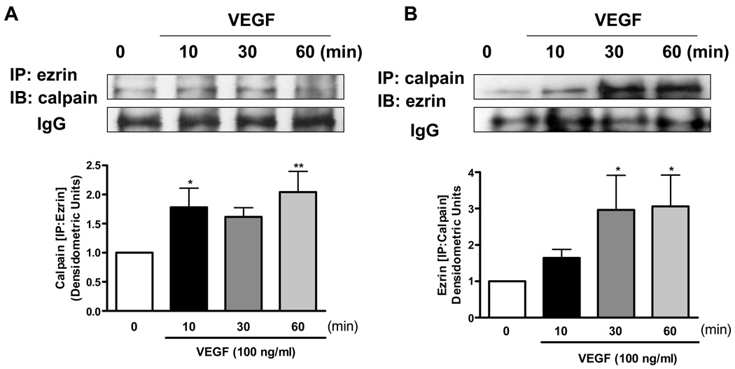
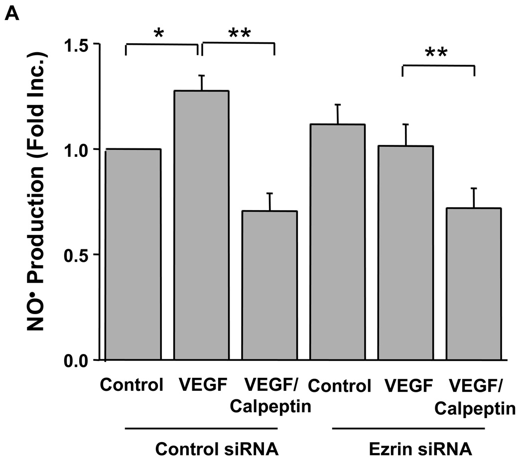
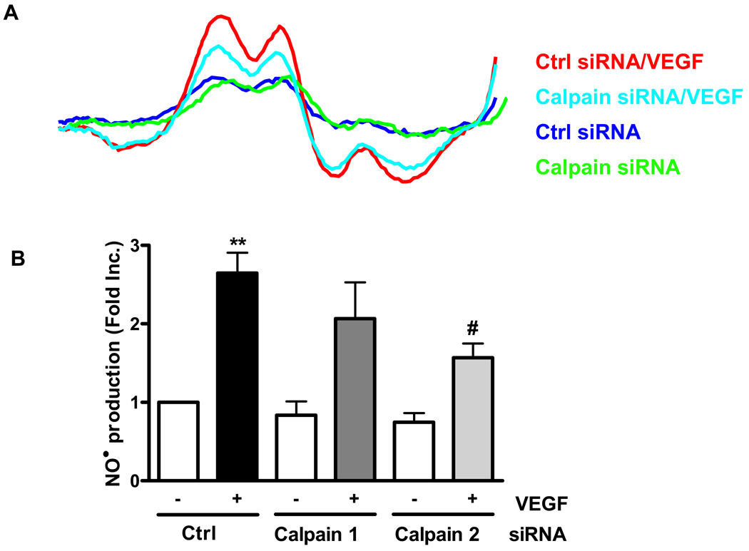
Calpain was recently reported to mediate vascular endothelial growth factor (VEGF)-induced angiogenesis. In the present study, we investigated detailed molecular mechanisms. VEGF (100 ng/mL) induced a marked increase in endothelial cell production of NO(*), specifically detected by electron spin resonance. This response was abolished by inhibition of calpain with N-acetyl-leucyl-leucyl-norleucinal (ALLN) or Calpeptin. Both also diminished membrane-specific calpain activation by VEGF, which was intriguingly attenuated by silencing ezrin with RNA interference. A rapid membrane colocalization of calpain and ezrin occurred as short as 10 minutes after VEGF stimulation. AKT, AMP-dependent kinase (AMPK), and endothelial nitric oxide synthase (eNOS)(s1179) phosphorylations in VEGF-stimulated endothelial cells were markedly enhanced, which were however significantly attenuated by either ALLN, Calpeptin, or ezrin small interfering RNA, as well as by Wortmannin or compound C (respectively for phosphatidylinositol 3-kinase [PI3K] or AMPK). The latter 3 also abolished VEGF induction of NO(*). These data indicate that AMPK and AKT are both downstream of PI3K and that AKT activation is partially dependent on AMPK. The interrelationship between AMPK and AKT, although known to be individually important in mediating VEGF activation of eNOS, is clearly characterized. Furthermore, AMPK/AKT/eNOS(s1179) was found downstream of a calpain/ezrin membrane interaction. These data no doubt provide new insights into the long mystified signaling gap between VEGF receptors and PI3K/AKT or AMPK-dependent eNOS activation. In view of the well-established significance of VEGF-dependent angiogenesis, these findings might have broad and important implications in cardiovascular pathophysiology.


Figures

Fig. 1
Fig. 1. Calpain plays a critical role in basal and VEGF-stimulated endothelial cell production of NO
Endothelial cells were pre-incubated with Calpeptin (10 µmol/L, 30 min) or ALLN (30 µmol/L, 30 min) prior to VEGF stimulation (100 ng/ml) and analysis of NO production using an electron spin resonance (ESR) spectrophotometer. A & B): Representative ESR spectra; C & D): Grouped densitometric data, Means±SEM, ANOVA, *p<0.05, **P<0.01
Fig. 2
Fig. 2. Colocalization of calpain and ezrin in response to VEGF
Endothelial cells cultured on coverslips were treated with VEGF for 10 min prior to immunostaining with fluorescent antibodies. Calpain and ezrin were visualized in green and red respectively, whereas nuclear DNA (in blue) was stained with Hoechst 33258. A & B): Representative subcellular localization of calpain and ezrin in un-stimulated and VEGF-stimulated endothelial cells.
Fig. 3
Fig. 3. Co-immunoprecipitation of calpain and ezrin
Endothelial cells were stimulated with 100 ng/ml of VEGF for indicated times. A &B): The cell lysates were subjected to immunoprecipitation with calpain or ezrin antibody, followed by immunoblotting with ezrin or calpain respectively. Grouped quantitative data are presented as Means±SEM (n=4), ANOVA; *p<0.05, **p<0.01
Fig. 4
Fig. 4. The role of ezrin in calpain-dependent endothelial NO production in response to VEGF
Proliferating endothelial cells were transfected with 25 nmol/L control or ezrin siRNA prior to Calpeptin (10 µmol/L, 30 min) and VEGF stimulation (100 ng/ml, 60 min). A): Grouped data of NO production are presented as Means±SEM, B): Representative western blot from cells transfected with ezrin siRNA, C & D): Ezrin mediated phosphorylation of eNOSs1179 and AKT in response to VEGF, ANOVA; *p<0.05, **p<0.01
Fig. 5
Fig. 5. Calpain-dependent phosphorylation of AKT and eNOS in response to VEGF
Endothelial cells were pre-treated with Calpeptin (10 µmol/L) or ALLN (30 µmol/L) for 30 min prior to VEGF stimulation (100 ng/ml, 10 min). A): AKT phosphorylation; B): eNOSs1179 phosphorylation, C): eNOSs116 phosphorylation, D): eNOSt497 phosphorylation. Representative western blots are shown with quantitative grouped data (Means±SEM, n=5), ANOVA, ***p<0.001 vs control, #p<0.5, ##p<0.01 vs VEGF.
Figure 6
Figure 6. The specific role of calpain-2 on endothelial NO bioavailability
BAECs were transfected with 25 nmol/L of control siRNA or 25 nmol/L of calpain 2 siRNA and then exposed to 100 ng/ml of VEGF. A): The bioavailable NO production was measured by using ESR. B): To investigate the role of calpain-2 in signaling mediating VEGF induced eNOS activation, western blotting was performed using indicated antibodies Grouped data are presented as mean ± SEM. ANOVA; *p<0.05, **p<0.01.
Fig. 7
Fig. 7. PI3K and AMPK are required for VEGF induction of NO
Endothelial cells were preincubated with an AMPK inhibitor compound C (20 µmol/L, 30min), or a PI3K antagonist wortmannin (100 nmol/L, 30 min), prior to VEGF stimulation (100 ng/ml, 60 min) and analysis of NO production. A): Representative ESR spectra; B): Grouped densitometric ESR data, Means±SEM (n=4), C): AMPK phosphorylation at thr 172 from BAECs pretreated with calpain inhibitors, calpeptin (10 µmol/L) or ALLN (30 µmol/L) for 30 min prior to VEGF (100 ng/ml, 10 min), D): Calpain activity from cells preincubated with a selective AMPK inhibitor, compound C (20 µmol/L, 30min), or H89 (10 µmol/L, 30 min) prior to 100 ng/ml of VEGF stimulation for 10 min. Grouped data is presented as means ± SEM (n=5). ANOVA; *p<0.05, **p<0.01 vs control
Fig. 8
Fig. 8. VEGF activation of eNOS is calpain, PI3K and AMPK-dependent
Endothelial cells were pre-treated with Compound C (20 µmol/L), wortmannin (100 nmol/L) or Calpeptin (10 µmol/L) for 30 min prior to VEGF stimulation (100 ng/ml, 10 min). Phosphorylation profiles of A): AMPK at thr 172, B): AKT at ser 473, and C): eNOS at ser 1179 are shown with quantitative grouped data (Means±SEM, n=4), ANOVA, *p<0.05 vs control, #p<0.05, ##p<0.01 vs VEGF.

References

    1. Khorchid A, Ikura M. How calpain is activated by calcium. Nat Struct Biol. 2002;9:239–241. - PubMed
    1. Tompa P, Emori Y, Sorimachi H, Suzuki K, Friedrich P. Domain III of calpain is a ca2+regulated phospholipid-binding domain. Biochem Biophys Res Commun. 2001;280:1333–1339. - PubMed
    1. Goll DE, Thompson VF, Li H, Wei W, Cong J. The calpain system. Physiol Rev. 2003;83:731–801. - PubMed
    1. Cuevas BD, Abell AN, Witowsky JA, Yujiri T, Johnson NL, Kesavan K, Ware M, Jones PL, Weed SA, DeBiasi RL, Oka Y, Tyler KL, Johnson GL. MEKK1 regulates calpain-dependent proteolysis of focal adhesion proteins for rear-end detachment of migrating fibroblasts. Embo J. 2003;22:3346–3355. - PMC - PubMed
    1. Kulkarni S, Saido TC, Suzuki K, Fox JE. Calpain mediates integrin-induced signaling at a point upstream of Rho family members. J Biol Chem. 1999;274:21265–21275. - PubMed

Publication types

MeSH terms