. 2009 Feb;132(Pt 2):544-57.
doi: 10.1093/brain/awn313. Epub 2008 Nov 27.

The course and outcome of unilateral intracranial arteriopathy in 79 children with ischaemic stroke

Affiliations

The course and outcome of unilateral intracranial arteriopathy in 79 children with ischaemic stroke

K P J Braun et al. Brain. 2009 Feb.

Abstract

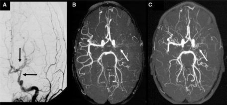
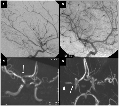
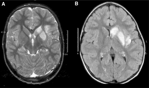
Arteriopathies are the commonest cause of arterial ischaemic stroke (AIS) in children. Repeated vascular imaging in children with AIS demonstrated the existence of a 'transient cerebral arteriopathy' (TCA), characterized by lenticulostriate infarction due to non-progressive unilateral arterial disease affecting the supraclinoid internal carotid artery and its proximal branches. To further characterize the course of childhood arteriopathies, and to differentiate TCA from progressive arterial disease, we studied the long-term evolution of unilateral anterior circulation arteriopathy, and explored predictors of stroke outcome and recurrence. From three consecutive cohorts in London, Paris and Utrecht, we reviewed radiological studies and clinical charts of 79 previously healthy children with anterior circulation AIS and unilateral intracranial arteriopathy of the internal carotid bifurcation, who underwent repeated vascular imaging. The long-term evolution of arteriopathy was classified as progressive or TCA. Clinical and imaging characteristics were compared between both groups. Logistic regression modelling was used to determine possible predictors of the course of arteriopathy, functional outcome and recurrence. After a median follow-up of 1.4 years, 5 of 79 children (6%) had progressive arteriopathy, with increasing unilateral disease or bilateral involvement. In the others (94%), the course of arteriopathy was classified as TCA. In 23% of TCA patients, follow-up vascular imaging showed complete normalization, the remaining 77% had residual arterial abnormalities, with improvement in 45% and stabilization in 32%. Stroke was preceded by chickenpox in 44% of TCA patients, and in none of the patients with progressive arteriopathies. Most infarcts were localized in the basal ganglia. In 14 (19%) of TCA patients, transient worsening of the arterial lesion was demonstrated before the arteriopathy stabilized or improved. Thirteen TCA patients (18%) had a recurrent stroke or TIA. Thirty TCA patients (41%) had a good neurological outcome, compared with none of the five patients with progressive arteriopathy. Arterial occlusion, moyamoya vessels and ACA involvement were more frequent in progressive arteriopathies. Cortical infarct localization was significantly associated with poor neurological outcome (OR 6.14, 95% CI 1.29-29.22, P = 0.02), while there was a trend for occlusive arterial disease to predict poor outcome (OR 3.00, 95% CI 0.98-9.23, P = 0.06). Progressive arteriopathy was associated with recurrence (OR 18.77, 95%CI 1.94-181.97, P = 0.01). The majority of childhood unilateral intracranial anterior circulation arteriopathies (94%) have a course that is consistent with TCA, in which transient worsening is common. Although the arterial inflammation probably causing TCA is 'transient', most children are left with permanent arterial abnormalities and residual neurological deficits.


Figures

Fig. 1
Fig. 1
Evolution of arterial abnormalities in a 6-year-old girl with typical TCA. (A) MRA at day 1 showing irregular stenosis of the A1 segment of the left ACA (left arrow) and M1 segment of the left MCA (right arrow). (B) Conventional contrast angiography 4 days after stroke illustrating irregular stenoses with string-of-beads sign (beading) involving the distal segment of the ICA, and the A1 and M1 and M2 segments of the ACA and MCA, respectively. (C) Complete resolution of arteriopathy (arrow) on MRA performed 15 months after stroke.
Fig. 2
Fig. 2
Another example of the course of arteriopathy in TCA. Conventional contrast angiography performed at 2 weeks (A) and 7 months (B) after stroke in a 7-year-old boy. (A) There is a long segmental irregular stenosis of the M1 of the MCA with beading at initial angiography. (B) Transient worsening of the disease with severe focal stenosis of the proximal M1 of the MCA (left arrow) and a long segmental stenosis of the M1 (right arrow). The beading, typical for the acute phase of TCA, is less prominent. Repeated angiography 22 months after stroke was identical, proving stabilization of arterial abnormalities (not shown).
Fig. 3
Fig. 3
Example of transient worsening in TCA, leading to complete MCA occlusion but subsequent improvement in an 8-year-old boy. (A) MRA shows a long segmental regular M1 stenosis (arrow) 1 day after stroke and ACA occlusion. (B) Three months later, there is complete occlusion of the origin of the MCA (arrow) and a tapered stenosis of the distal ICA. (C) Twenty months after stroke, MRA has almost normalized, with a residual focal MCA stenosis (arrow) and patent (hypoplastic?) ACA.
Fig. 4
Fig. 4
Another example of occlusive TCA with subsequent normalization. (A) Conventional angiography 2 weeks after basal ganglia stroke in a 7-year-old girl, showing a long tight distal ICA stenosis (horizontal arrow) and occlusion of the proximal MCA (vertical arrow). (B) Eight years later, there is complete normalization of arteriopathic abnormalities (the arrow points at a flow artefact in the A1 of the ACA).
Fig. 5
Fig. 5
Occlusive stabilizing unilateral TCA in a 3-year-old boy who presented with three TIAs and a basal ganglia infarct on MRI. (A) Conventional angiography at 11 days after stroke shows an irregular distal ICA stenosis (horizontal arrow) and a proximal MCA occlusion with the development of some abnormal moyamoya lenticulostriate collaterals (vertical arrow). MRA at 3 months (B) and 3 years (C) after stroke shows a persisting but unchanged MCA occlusion with some abnormal moyamoya collaterals (arrows). There is no contralateral involvement in time.
Fig. 6
Fig. 6
Progressive arteriopathy in a girl who initially presented with an ischaemic stroke in the left basal ganglia and loss of flow voids in the left ICA and proximal MCA on MRA, 8 days after stroke. She was treated with aspirin and was left with a moderate right hemiparesis and some comprehension and word-finding difficulties. Conventional angiography at 3 months after stroke showed vasculopathy affecting the left side (A) but the right was normal (B). Further MRA 9 months later (C) suggested abnormality of the right proximal ACA (arrow), but the findings were interpreted cautiously in view of the image quality and it was not considered ethical to repeat the conventional arteriography. Three months later, she had a contralateral infarct on MRI, involving the right basal ganglia and insular region (not shown). MRA (D), confirmed by conventional angiography, then showed bilateral arteriopathy with proximal stenosis of the right MCA (arrow) and a more distal occlusion of the right M1 segment (arrow head), as well as irregular stenosis of the left MCA.
Fig. 7
Fig. 7
Typical examples of infarct localization in TCA, including the head of the caudate nucleus, the lentiform nucleus, some involvement but relative sparing of the internal capsule and some extension in adjacent gray or white matter. (A) T2-weighted MRI in a 7-year-old boy. (B) FLAIR MRI in a 6-year-old girl.

References

    1. Askalan R, Laughlin S, Mayank S, Chan A, MacGregor D, Andrew M, et al. Chickenpox and stroke in childhood: a study of frequency and causation. Stroke. 2001;32:1257–62. - PubMed
    1. Aviv RI, Benseler SM, Silverman ED, Tyrrell PN, deVeber G, Tsang LM, et al. MR imaging and angiography of primary CNS vasculitis of childhood. AJNR Am J Neuroradiol. 2006;27:192–9. - PMC - PubMed
    1. Benseler SM, deVeber G, Hawkins C, Schneider R, Tyrrell PN, Aviv RI, et al. Angiography-negative primary central nervous system vasculitis in children: a newly recognized inflammatory central nervous system disease. Arthritis Rheum. 2005;52:2159–67. - PubMed
    1. Benseler SM, Silverman E, Aviv RI, Schneider R, Armstrong D, Tyrrell PN, et al. Primary central nervous system vasculitis in children. Arthritis Rheum. 2006;54:1291–7. - PubMed
    1. Berger TM, Caduff JH, Gebbers JO. Fatal varicella-zoster virus antigen-positive giant cell arteritis of the central nervous system. Pediatr Infect Dis J. 2000;19:653–6. - PubMed

Publication types

MeSH terms