

Case Reports
doi: 10.3348/kjr.2008.9.6.550.
Duplication of the extrahepatic bile duct in association with choledocholithiasis as depicted by MDCT
Affiliations
- PMID: 19039272
- PMCID: PMC2627237
- DOI: 10.3348/kjr.2008.9.6.550
Item in Clipboard
Case Reports
Duplication of the extrahepatic bile duct in association with choledocholithiasis as depicted by MDCT
Korean J Radiol.
2008 Nov-Dec.
Abstract
We report here on an extremely rare case of duplicated extrahepatic bile ducts that was associated with choledocholithiasis, and this malady was visualized by employing the minimum intensity projection images with using multi-detector row CT. The presence of duplicated extrahepatic bile ducts with a proximal communication, and the ducts were joined distally and they subsequently formed a single common bile duct, has not been previously reported.
Figures
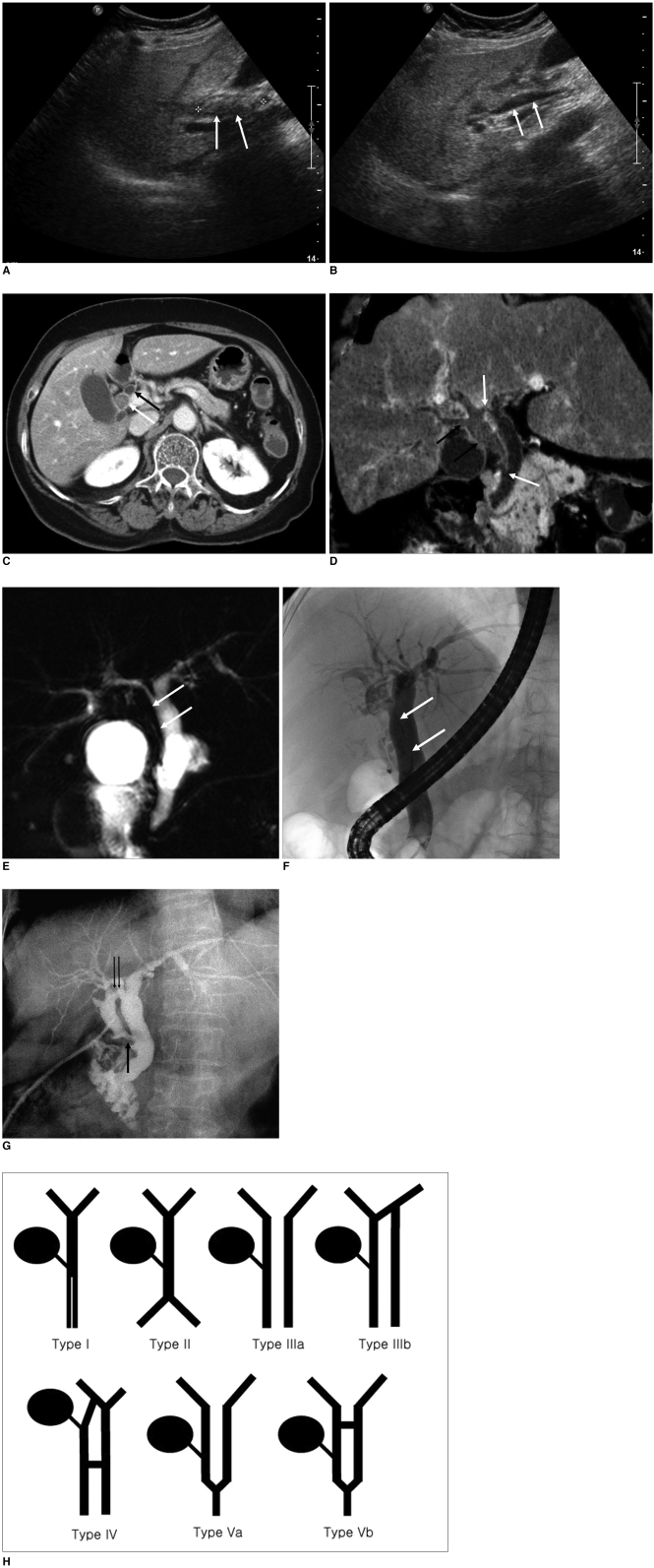
81-year-old woman with duplication of extrahepatic bile duct. A. Sagittal sonography shows dilated extrahepatic bile duct (white arrows) filled with shadowing echogenic materials (between electronic calipers). B. On more medial view, another extrahepatic bile duct is seen (white arrows). No intraluminal lesion is found in this duct. C. Contrast-enhanced axial CT scan shows two separate extrahepatic bile ducts (arrows), one of which was filled with stones (white arrow). D. Minimum intensity projection image shows proximal and distal unions of duplicated extrahepatic bile ducts (white arrows). Also note stones in laterally located extrahepatic bile duct and right intrahepatic bile ducts. These ducts show higher attenuation than that of water (black arrows). E. MR cholangiogram shows complete signal void in laterally located extrahepatic bile duct due to multiple impacted stones (arrows). F. Balloon cholangiography with forceful injection of contrast material shows another stone-filled extrahepatic bile duct (white arrows). G. Operative cholangiography shows same anatomical details of extraheptic bile ducts as those depicted by CT and endoscopic retragrade cholangiography images. The black arrows indicate proximal (double arrows) and distal (single arrow) unions of double extrahepatic bile ducts. H. Modified classification system of duplicated extrahepatic bile duct as proposed by Choi et al. (5).
References
-
- Teilum D. Double common bile duct. Case report and review. Endoscopy. 1986;18:159–161. - PubMed
-
- Yamashita K, Oka Y, Urakami A, Iwamoto S, Tsunoda T, Eto T. Double common bile duct: a case report and a review of the Japanese literature. Surgery. 2002;131:676–681. - PubMed
-
- Leung B, Lai P, Chan A, Chan YL, Lau WY. A variant of an accessory common bile duct. Endoscopy. 2000;32:728–730. - PubMed
-
- Balbinot RA, Gobbato A, Balbinot SS, Mendonca L, Tefilli N, Lain VV, et al. Double bile duct with ectopic drainage into stomach. Gastrointest Endosc. 2004;60:661–662. - PubMed
-
- Choi E, Byun JH, Park BJ, Lee MG. Duplication of the extrahepatic bile duct with anomalous union of the pancreaticobiliary ductal system revealed by MR cholangiopancreatography. Br J Radiol. 2007;80:E150–E154. - PubMed
Publication types
MeSH terms
LinkOut - more resources
Full Text Sources
