

Aptamers selected against the unglycosylated EGFRvIII ectodomain and delivered intracellularly reduce membrane-bound EGFRvIII and induce apoptosis
- PMID: 19040357
- PMCID: PMC3816755
- DOI: 10.1515/BC.2009.022
Aptamers selected against the unglycosylated EGFRvIII ectodomain and delivered intracellularly reduce membrane-bound EGFRvIII and induce apoptosis
Abstract
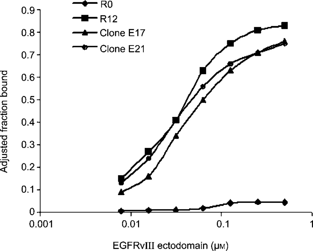
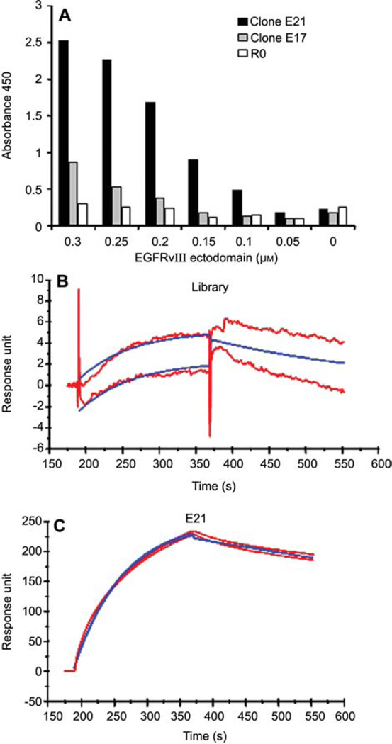
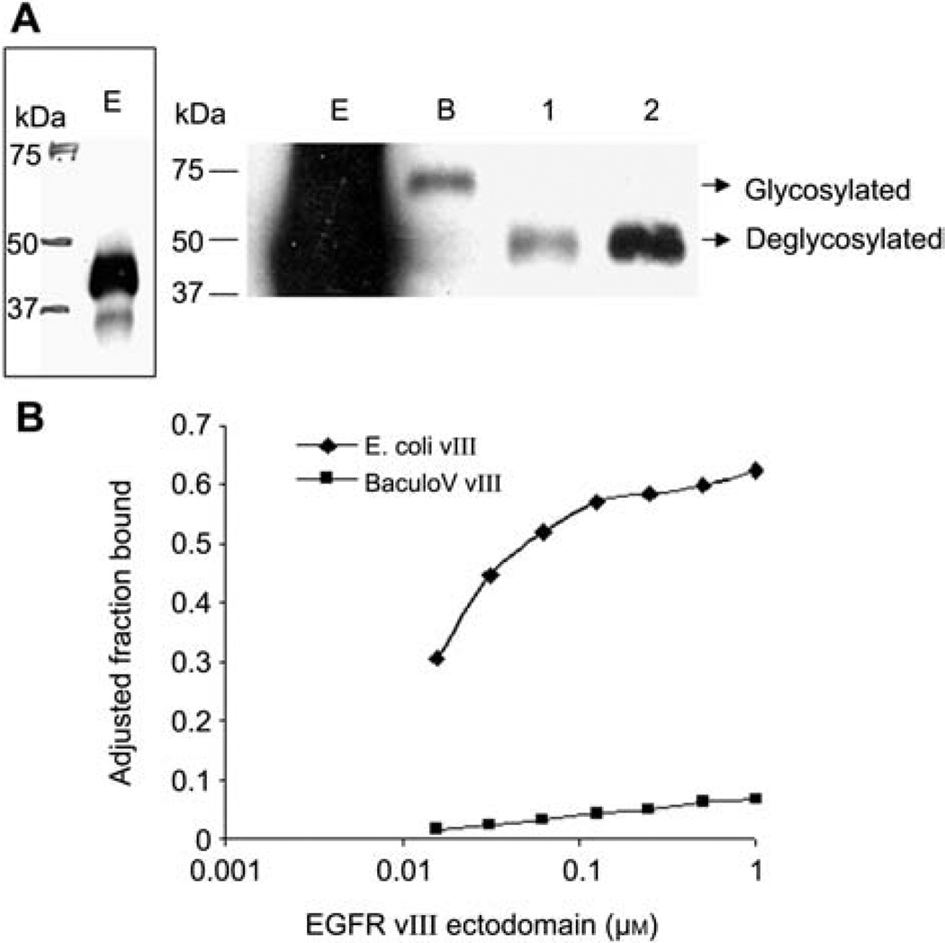
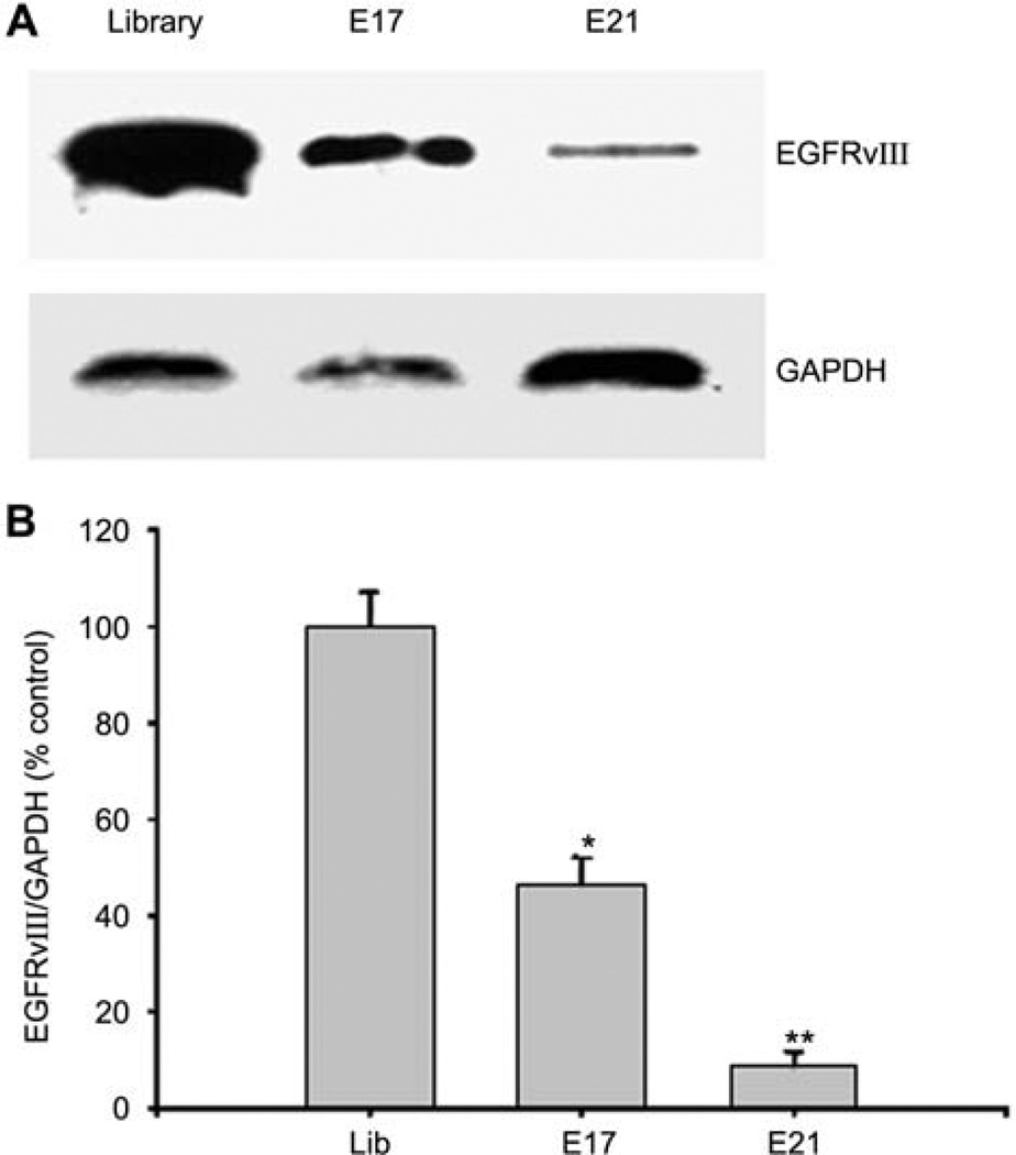
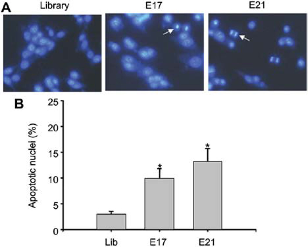
Epidermal growth factor receptor variant III (EGFRvIII) is a glycoprotein uniquely expressed in glioblastoma, but not in normal brain tissues. To develop targeted therapies for brain tumors, we selected RNA aptamers against the histidine-tagged EGFRvIII ectodomain, using an Escherichia coli system for protein expression and purification. Representative aptamer E21 has a dissociation constant (Kd) of 33x10(-9) m, and exhibits high affinity and specificity for EGFRvIII in ELISA and surface plasmon resonance assays. However, selected aptamers cannot bind the same protein expressed from eukaryotic cells because glycosylation, a post-translational modification present only in eukaryotic systems, significantly alters the structure of the target protein. By transfecting EGFRvIII aptamers into cells, we find that membrane-bound, glycosylated EGFRvIII is reduced and the percentage of cells undergoing apoptosis is increased. We postulate that transfected aptamers can interact with newly synthesized EGFRvIII, disrupt proper glycosylation, and reduce the amount of mature EGFRvIII reaching the cell surface. Our work establishes the feasibility of disrupting protein post-translational modifications in situ with aptamers. This finding is useful for elucidating the function of proteins of interest with various modifications, as well as dissecting signal transduction pathways.
Figures
References
-
- Baselga J. Why the epidermal growth factor receptor? The rationale for cancer therapy. Oncologist. 2002;7(Suppl. 4):2–8. - PubMed
-
- Batra SK, Castelino-Prabhu S, Wikstrand CJ, Zhu X, Humphrey PA, Friedman HS, Bigner DD. Epidermal growth factor ligand-independent, unregulated, cell-transforming potential of a naturally occurring human mutant EGFRvIII gene. Cell Growth Differ. 1995;6:1251–1259. - PubMed
-
- Bunka DH, Stockley PG. Aptamers come of age – at last. Nat. Rev. Microbiol. 2006;4:588–596. - PubMed
-
- Durand G, Seta N. Protein glycosylation and diseases: blood and urinary oligosaccharides as markers for diagnosis and therapeutic monitoring. Clin. Chem. 2000;46:795–805. - PubMed
Publication types
MeSH terms
Substances
Grants and funding
LinkOut - more resources
Full Text Sources
Other Literature Sources
Research Materials