

Analysis of molecular determinants of PRL-3
- PMID: 19040419
- PMCID: PMC4516473
- DOI: 10.1111/j.1582-4934.2008.00591.x
Analysis of molecular determinants of PRL-3
Abstract
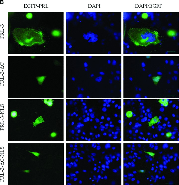
In order to analyse whether a C-terminal polybasic sequence represents a nuclear localization signal (NLS) we obtained several truncated and mutant forms of protein of regerating liver (PRL)-3 and evaluated their subcellular localization as compared to the wild-type form. Our results invalidate the hypothesis that this is an NLS. We also analysed the influence of the C- and N-terminal residues on the phosphatase activity of PRL-3. Our results provide in vitro evidence that the C-terminal CAAX motif, besides directing the protein farnesylation, plays an additional regulatory role by inhibiting the catalytic efficiency of PRL-3. Taking into account the results we obtained, as well as reported data, we propose a hypothetical molecular mechanism for the nucleocytoplasmic localization and transfer of PRL-3.
Figures


Similar articles
-
Regulatory mechanisms of phosphatase of regenerating liver (PRL)-3.Biochem Soc Trans. 2016 Oct 15;44(5):1305-1312. doi: 10.1042/BST20160146. Biochem Soc Trans. 2016. PMID: 27911713 Free PMC article. Review.
-
Enzyme activity of phosphatase of regenerating liver is controlled by the redox environment and its C-terminal residues.Biochemistry. 2009 May 26;48(20):4262-72. doi: 10.1021/bi900241k. Biochemistry. 2009. PMID: 19341304 Free PMC article.
-
Thioredoxin-related protein 32 (TRP32) specifically reduces oxidized phosphatase of regenerating liver (PRL).J Biol Chem. 2013 Mar 8;288(10):7263-70. doi: 10.1074/jbc.M112.418004. Epub 2013 Jan 28. J Biol Chem. 2013. PMID: 23362275 Free PMC article.
-
PRL-3 down-regulates PTEN expression and signals through PI3K to promote epithelial-mesenchymal transition.Cancer Res. 2007 Apr 1;67(7):2922-6. doi: 10.1158/0008-5472.CAN-06-3598. Cancer Res. 2007. PMID: 17409395
-
Molecular mechanisms of the PRL phosphatases.FEBS J. 2013 Jan;280(2):505-24. doi: 10.1111/j.1742-4658.2012.08565.x. Epub 2012 Apr 10. FEBS J. 2013. PMID: 22413991 Review.
Cited by
-
PRL-3 promotes telomere deprotection and chromosomal instability.Nucleic Acids Res. 2017 Jun 20;45(11):6546-6571. doi: 10.1093/nar/gkx392. Nucleic Acids Res. 2017. PMID: 28482095 Free PMC article.
-
PRL-3 promotes the peritoneal metastasis of gastric cancer through the PI3K/Akt signaling pathway by regulating PTEN.Oncol Rep. 2016 Oct;36(4):1819-28. doi: 10.3892/or.2016.5030. Epub 2016 Aug 23. Oncol Rep. 2016. PMID: 27572739 Free PMC article.
-
Regulatory mechanisms of phosphatase of regenerating liver (PRL)-3.Biochem Soc Trans. 2016 Oct 15;44(5):1305-1312. doi: 10.1042/BST20160146. Biochem Soc Trans. 2016. PMID: 27911713 Free PMC article. Review.
-
Identification of proteins suppressing the functions of oncogenic phosphatase of regenerating liver 1 and 3.Exp Ther Med. 2016 Nov;12(5):2974-2982. doi: 10.3892/etm.2016.3722. Epub 2016 Sep 20. Exp Ther Med. 2016. PMID: 27882103 Free PMC article.
-
Downregulation of phosphatase of regenerating liver-3 is involved in the inhibition of proliferation and apoptosis induced by emodin in the SGC-7901 human gastric carcinoma cell line.Exp Ther Med. 2012 Jun;3(6):1077-1081. doi: 10.3892/etm.2012.516. Epub 2012 Mar 15. Exp Ther Med. 2012. PMID: 22970020 Free PMC article.
References
-
- Stephens BJ, Han H, Gokhale V, et al. PRL phosphatases as potential molecular targets in cancer. Mol Cancer Ther. 2005;4:1653–61. - PubMed
-
- Zeng Q, Hong W, Tan YH. Mouse PRL-2 and PRL-3, two potentially prenylated protein tyrosine phosphatases homologous to PRL-1. Biochem Biophys Res Commun. 1998;244:421–7. - PubMed
-
- Saha S, Bardelli A, Buckhaults P, et al. A phosphatase associated with metastasis of colorectal cancer. Science. 2001;294:1343–6. - PubMed
Publication types
MeSH terms
Substances
LinkOut - more resources
Full Text Sources
