. 2008 Dec;41(6):936-59.
doi: 10.1111/j.1365-2184.2008.00566.x.

Role of alpha7-nicotinic acetylcholine receptor in human non-small cell lung cancer proliferation

Affiliations

Role of alpha7-nicotinic acetylcholine receptor in human non-small cell lung cancer proliferation

L Paleari et al. Cell Prolif. 2008 Dec.

Erratum in

  • Cell Prolif. 2009 Aug;42(4):568
  • Cell Prolif. 2009 Feb;42(1):122

Retraction in

Abstract

Objectives: Lung cancer is the most common cause of cancer death in the world. Cigarette smoking represents the major risk factor. Nicotine, an active component of cigarettes, can induce cell proliferation, angiogenesis and apoptosis resistance. All these events are mediated through the nicotinic acetylcholine receptor (nAChR) expressed on lung cancer cells. We speculate that new insights into the pathophysiological roles of nAChR may lead to new therapeutic avenues to reduce non-small cell lung cancer (NSCLC) tumour growth.

Materials and methods: Human samples of NSCLC, cell lines and mouse models were utilized in Western blotting, reverse transcriptase polymerase chain reaction and apoptosis studies.

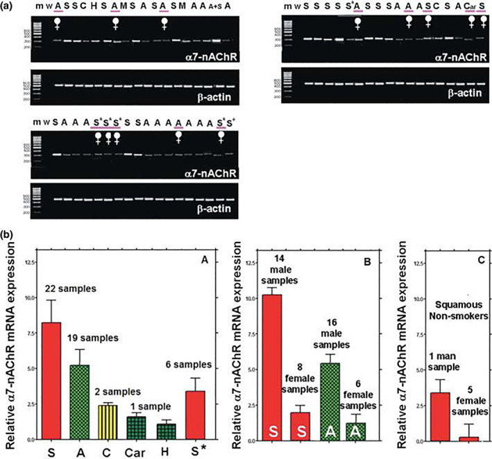
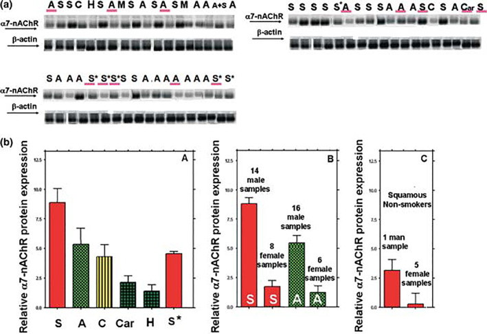
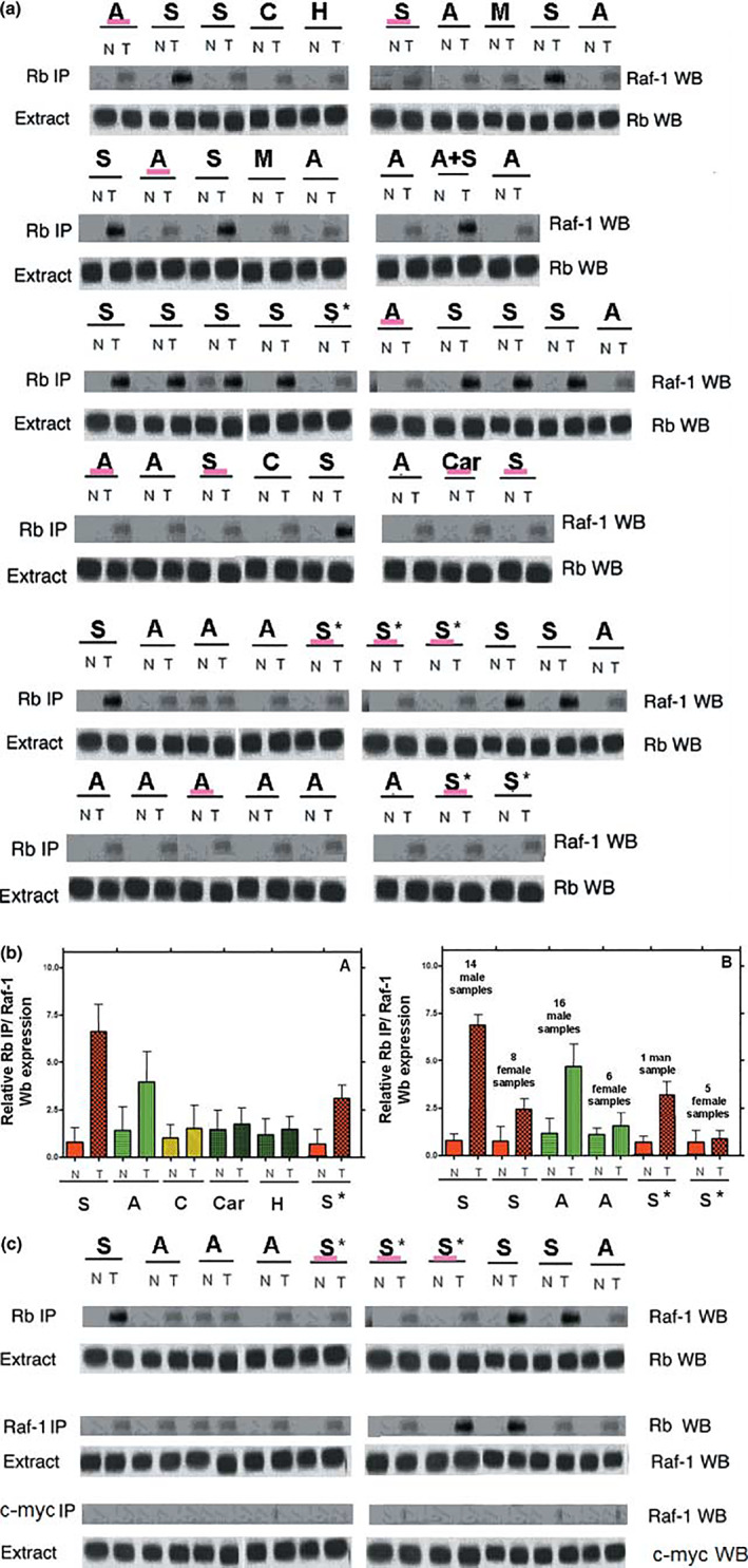
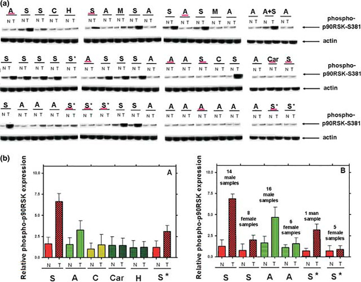
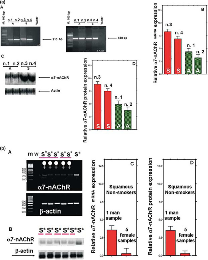
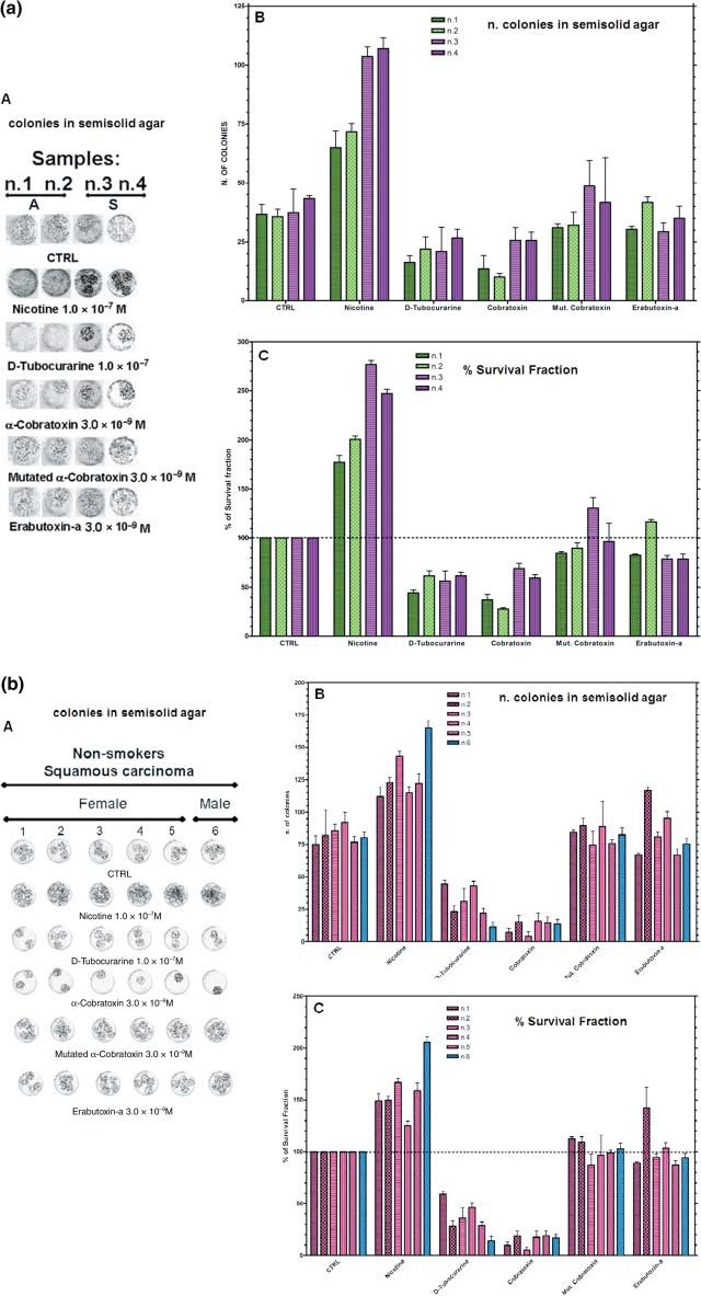
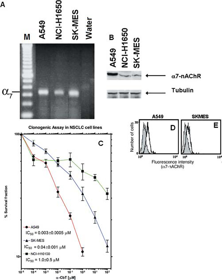
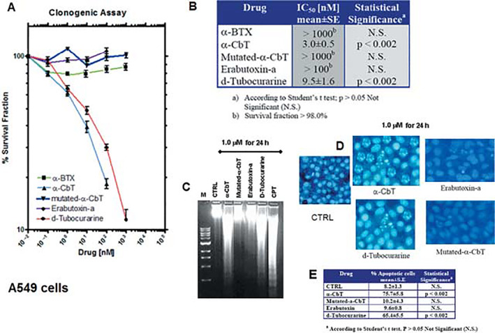
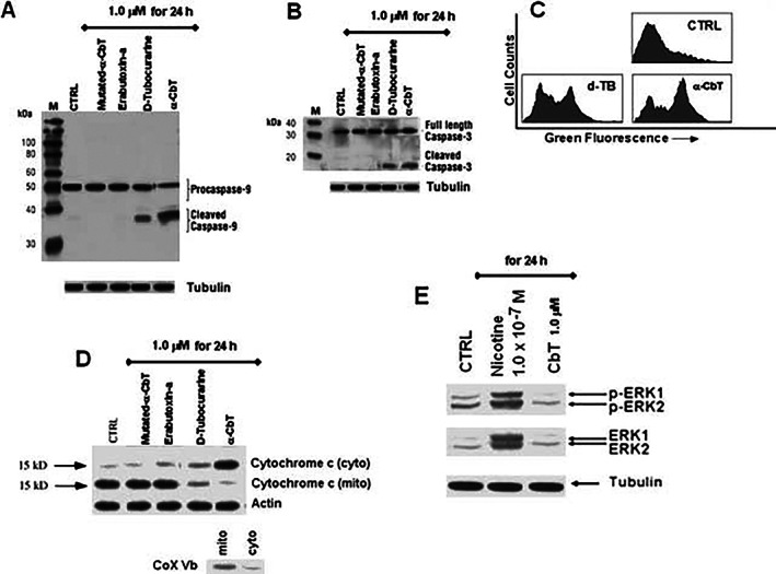
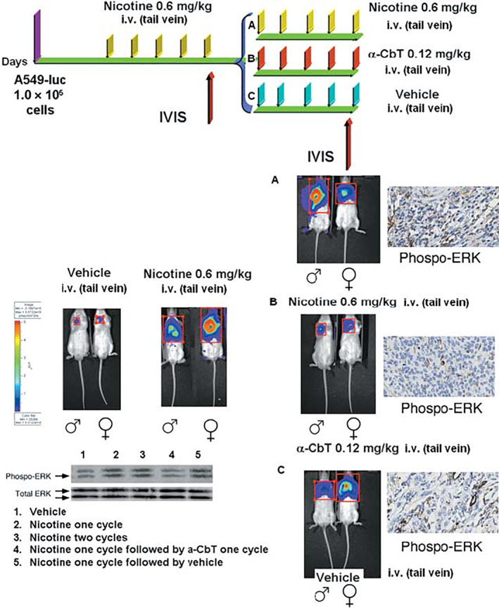
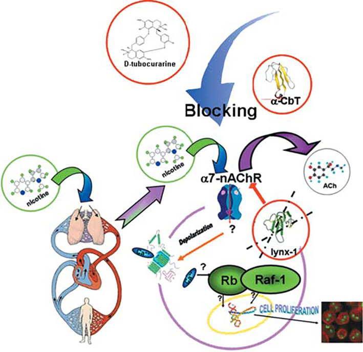
Results: Human NSCLC tissues expressed alpha7-nAChR. This expression was higher in smoking patients with squamous carcinomas than those with adenocarcinomas and in male smoking patients than in females. All the data support the hypothesis that major expression of alpha7-nAChR is related to major activation of the Rb-Raf-1/phospho-ERK/phospho-p90RSK pathway. alpha7-nAChR antagonists, via mitochondria associated apoptosis, inhibited proliferation of human NSCLC primary and established cells. Nicotine stimulates tumour growth in a murine model, A549 cells orthotopically grafted. The effects of nicotine were associated with increases in phospho-ERK in tumours. Proliferation effects of nicotine could be blocked by inhibition of alpha7-nAChR by the high affinity ligand alpha-cobratoxin.

Conclusion: These results showed that alpha7-nAChR plays an important role in NSCLC cell growth and tumour progression as well as in cell death.


Figures

Figure 1
Figure 1
Expression of α7‐nAChR in human NSCLC tissue samples. (a) RT‐PCR analysis showing expression of α7‐nAChR mRNA. m, marker; w, water. (b) Densitometric analysis. Each bar represents the mean ± standard error of at least three independent experiments. Squamous carcinomas (S), squamous carcinomas from non‐smokers (S*), adenocarcinomas (A), adeno/squamous carcinoma (A + S), large cell carcinomas (C), lung cancer metastasis (M) and samples of non‐neoplastic/malignant tissues such as carnification (Car) and pulmonary chondroid hamartoma (H). Panel A: total samples. Panel B: gender and histology classification partition. P < 0.002 (males versus females) for squamous carcinoma and for adenocarcinoma; P < 0.002 for male squamous carcinoma versus male adenocarcinoma; P > 0.05 for female squamous carcinoma versus female adenocarcinoma. Panel C: gender and histology classification partition for former non‐smokers. P < 0.05 (male versus female) for squamous carcinoma; according to two‐tailed paired Student's t‐test, Spearman correlation test and non‐parametric Mann–Whitney U‐test.
Figure 2
Figure 2
Expression of α7‐nAChR in human NSCLC tissue samples. (a) Western blot analysis for α7‐nAChR protein. (b) Densitometric analysis. Each bar represents the mean ± standard error of at least three independent experiments. Squamous carcinomas (S), squamous carcinomas from non‐smokers (S*), adenocarcinomas (A), adeno/squamous carcinoma (A + S), large cell carcinomas (C), lung cancer metastasis (M) and samples of non‐neoplastic/malignant tissues carnification (Car) and pulmonary chondroid hamartoma (H). Panel A: total samples. Panel B: gender and histology classification partition. P < 0.002 (male versus female) for squamous carcinoma and for adenocarcinoma; P < 0.002 for male squamous carcinoma versus male adenocarcinoma; P > 0.05 for females squamous carcinoma versus female adenocarcinoma. Panel C: gender and histology classification partition for former non smokers. P < 0.05 (male versus female) for squamous carcinoma; according to two‐tailed paired Student's t‐test, Spearman correlation test and non‐parametric Mann–Whitney U‐test.
Figure 3
Figure 3
Rb–Raf‐1 interactions in human NSCLC tissue samples. (a) Co‐immunoprecipitation of Raf‐1 with Rb; whole‐cell extracts were immunoprecipitated with antibodies to Rb. Western blot analysis revealed the presence of Raf‐1 in Rb. An equivalent amount of samples extracts were used for immunoprecipitation. (b) Densitometric analysis, each bar represents the mean ± standard error of at least three independent experiments. Panel A: total samples. Panel B: gender and histological partition. N, unaffected tissue; T, tumour tissue. P < 0.002 (T versus N) for squamous carcinoma and for adenocarcinoma: P = 0.05 for large carcinoma and for squamous carcinoma from non‐smokers; P > 0.05 (not significant) for carnification and pulmonary chondroid hamartoma according to Student's t‐test, Spearman correlation test and non‐parametric Mann–Whitney U‐test. (c) Whole‐cell extracts were immunoprecipitated with a control (c‐myc) or a Raf‐1 antibody. The immunoprecipitates were examined for the presence of Rb (top) or c‐myc (bottom) by Western blotting. The first two lines represent some experiments already reported in Fig. 3a.
Figure 4
Figure 4
Phosphorylated levels of p90SSK (Ser 381) in human NSCLC tissue samples. (a) Western blotting. (b) Densitometric analyses. Panel A: total samples. Panel B: gender and histological partition. N, unaffected tissue; T, tumour tissue. P < 0.002 (T versus N) for squamous carcinoma and for adenocarcinoma: P = 0.05 for large carcinoma and for squamous carcinoma from non‐smokers; P > 0.05 (not significant) for carnification and pulmonary chondroid hamartoma according to Student's t‐test, Spearman correlation test and non‐parametric Mann–Whitney U‐test.
Figure 5
Figure 5
Effects of nicotine or nAChR antagonists, namely, d‐tubocurarine, α‐CbT, mutated α‐CbT or Erabutoxin‐a, on primary culture obtained from biopsies of different patients. (a) Expression of mRNA (panel A) and relative proteins for α7‐nAChR (panel C), and densitometric analysis (panels B and D) from four heavy smoker biopsies. Each bar represents the mean ± standard error (SE) of at least three independent experiments. P < 0.002 for squamous carcinoma (S) versus adenocarcinoma (A); P > 0.05 (not significant) for sample 1 versus sample 2 and for sample 3 versus sample 4, according to two‐tailed paired Student's t‐test, Spearman correlation and non‐parametric Mann–Whitney U‐test. (b) Expression of mRNA (panel A) and relative proteins for α7‐nAChR (panel B), and densitometric analysis (panels C and D) from six non‐smokers squamous carcinoma (S*) biopsies. Each bar represents the mean ± SE of at least three independent experiments. P < 0.002 for male versus female, according to two‐tailed paired Student's t‐test, Spearman correlation and non‐parametric Mann–Whitney U‐test.
Figure 6
Figure 6
Effects of nicotine or nAChR antagonists, namely, d‐tubocurarine, α‐CbT, mutated α‐CbT or Erabutoxin‐a, on primary culture obtained from biopsies of different patients. Clonogenic activity in semisolid agar. (a) Primary cells from four heavy smokers’ biopsies. Cells (5.0 × 10⁵) were plated on 6‐multiwells plates. Drugs were administered three times a week for 21 weeks. Panel A: cell colonies in semisolid agar. Panel B: no. of colonies. Panel C: % survival fraction. P < 0.002 for eac versus h sample treated with nicotine, d‐tubocurarine or α‐CbT (samples from 1 to 4) versus their own control cells, P > 0.05 (not significant) for each sample treated with Erabutoxin‐a or mutated α‐CbT (1 to 4) versus their own control cells. P < 0.002 for (3, 4) treated squamous samples with nicotine, d‐tubocurarine or α‐CbT versus treated adenocarcinoma samples (1, 2) with nicotine, d‐tubocurarine or α‐CbT, according to two‐tailed paired Student's t‐test and non‐parametric Mann–Whitney U‐test. (b) Primary cells from six non‐smokers squamous carcinoma patients. Clonogenic activity in semisolid agar. Cells (5.0 × 10⁵) were plated on 6‐multiwells plates. Panel A: cell colonies in semisolid agar. Panel B: no. of colonies. Panel C: % survival fraction. Drugs were administered three times a week for 21 weeks. P < 0.002 for each sample treated with nicotine, d‐tubocurarine or α‐CbT (1 to 6) their own control cells, P > 0.05 (not significant) for each sample treated with Erabutoxin‐a or mutated α‐CbT (1 to 6) versus their own control cells, according to two‐tailed paired Student's t‐test and nonparametric Mann–Whitney U‐test.
Figure 7
Figure 7
Effects of the high‐affinity nAChR ligand α‐CbT on NSCLC cell lines. Panel A and panel B: expression of mRNA (panel A) and relative proteins (panel B) for α‐7nAChR by A549, SK‐MES and NCI‐H1650 NSCLC cell lines. Panel C: survival fraction. One thousand cells were plated on 6‐multiwells plates. Different concentrations of α‐CbT were administered three times a week for 21 weeks. Panel (D) A549 and panel E (SK‐MES) binding experiments using α‐CbT‐FITC.
Figure 8
Figure 8
Effects of nAChR antagonists, namely, d‐tubocurarine, α‐CbT, mutated‐α‐CbT, α‐BTX or Erabutoxin‐a, on NSCLC cell line A549. Panel A: survival fraction; panel B: IC50 values; panel C: apoptosis evaluation by gel ladder (M, marker; CPT, campthotecin‐positive control); panel D: apoptosis evaluation by scoring DAPI‐stained cells. Panel E: % of apoptotic cells of DAPI‐stained cells.
Figure 9
Figure 9
Characterization of apoptosis induced by d‐tubocurarine, α‐CbT, mutated‐α‐CbT or Erabutoxin‐a on NSCLC cell line A549. Panel A: cleavage of procaspase‐9. Panel B: cleavage of procaspase‐3. Panel C: mitochondrial potential. Panel D: cytochrome c release. Panel E: ERK phosphorylation (Western blotting), nicotine represented positive control.
Figure 10
Figure 10
In vivo experiments. Top, schedule of treatment: at day zero orthotopically cell implantation (1.0 × 10⁵), after 48 h of recovery, at day 4, starting of treatment with nicotine for 5 consecutive days, after 48 h of recovery, at day 10, randomization and starting treatment with different drugs for additional 5 days. Final evaluation at day 14. Bottom: mice treated with vehicle alone for five times, mice treated with nicotine for five times, then nicotine‐treated animals received nicotine for additional five times, a‐CbT for additional five times, or vehicle alone for additional 5 days. Immunohistochemistry and Western blotting of phospho‐ERK.
Figure 11
Figure 11
Possible mechanism of action of α‐7nAChR agonist or antagonist on NSCLC growth.

Similar articles

Cited by

References

    1. Antil‐Delbeke S, Gaillard C, Tamiya T (2000) Molecular determinants by which a long chain toxin from snake venom interacts with the neuronal alpha7‐nicotinic acetylcholine receptor. J. Biol. Chem. 275, 29594–29601. - PubMed
    1. Arredondo J, Chernyavsky AI, Grando SA (2006a) Nicotinic receptors mediate tumorigenic action of tobacco‐derived nitrosamines on immortalized oral epithelial cells. Cancer Biol. Ther. 5, 511–517. - PubMed
    1. Arredondo J, Chernyavsky AI, Grando SA (2006b) The nicotinic receptor antagonists abolish pathobiologic effects of tobacco‐derived nitrosamines on BEP2D cells. J. Cancer Res. Clin. Oncol. 132, 653– 663. - PubMed
    1. Battersby AR, Hodson HF (1968) Alkaloids of calabash curare and Strychnos species. In: Manske RHF, ed. The Alkaloids, Chemistry and Physiology, Vol. 11. Academic Press, New York, pp. 189–204.
    1. Boffetta P, Agudo A, Ahrens W, Benhamou E, Benhamou S, Darby SC, Ferro G, Fortes C, Gonzalez CA, Jockel KH, Krauss M, Kreienbrock L, Kreuzer M, Mendes A, Merletti F, Nyberg F, Pershagen G, Pohlabeln H, Riboli E, Schmid G, Simonato L, Tredaniel J, Whitley E, Wichmann HE, Winck C, Zambon P, Saracci R (1998) Multicenter case control study of exposure to environmental tobacco smoke and lung cancer in Europe. J. Natl Cancer Inst. 90, 1440–1450. - PubMed

Publication types

MeSH terms