

A spindle cell carcinoma presenting with osseous metaplasia in the gingiva: a case report with immunohistochemical analysis
- PMID: 19040765
- PMCID: PMC2627831
- DOI: 10.1186/1746-160X-4-28
A spindle cell carcinoma presenting with osseous metaplasia in the gingiva: a case report with immunohistochemical analysis
Abstract
Background: Spindle cell carcinoma (SpCC) is a rare, high malignant variant of squamous cell carcinoma (SCC), which shows biphasic proliferation of conventional SCC component and malignant spindle shape cells with sarcomatous appearance.
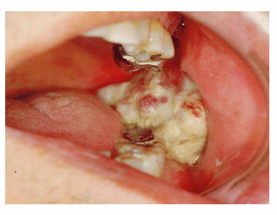
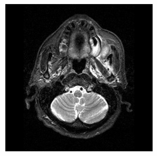
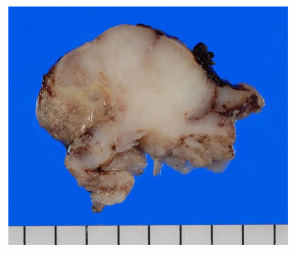
Methods: A case of Spindle cell carcinoma with bone-like calcified materials, occurring at the mandibular molar region of 71-years-old Japanese male patient was presented with gross finding, histological findings and MRI image. To identify the characteristics of the bone-like materials, immunohistochemistry were performed.
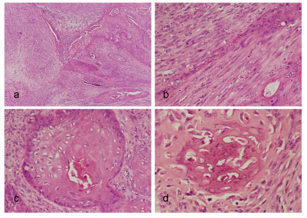
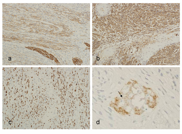
Results: Histologically, the cancer cells were composed of spindle cells and epithelial cells which form nests with prominent keratinization. Histological findings showed typical histology of the SpCC, however, as an uncommon finding, spatters of calcified, bone-like materials were observed in between the cancer cells. Immunohistochemistry revealed that cancer cells were positive for cytokeratins and vimentin to a varying degree and negative for Desmin, S-100, Osteopontin, BMP-2 or BMP-4. These findings implied that the calcified materials were formed by metaplasia of the stromal cells.
Discussion: Bone-like materials formation by osseous and/or cartilaginous metaplasia of the stroma in the carcinoma has been reported. However, the detailed mechanism of these metaplasia and affection on the clinical feature, prognosis and therapies are not well established. In summary, we presented an unique case of SpCC, which has not been described in the literature.
Figures
References
-
- Takata T, Ito H, Ogawa I, Miyauchi M, Ijuhin N, Nikai H. Spindle cell squamous carcinoma of the oral region. An immunohistochemical and ultrastractual study on the histogenesis and differential diagnosis with a clinicopathological analysis of six cases. Virchows Arch. 1991;419:177–182. doi: 10.1007/BF01626345. - DOI - PubMed
-
- Torenbeek R, Hermsen MA, Meijer GA, Baak JP, Meijer CJ. Analysis by comparative genomic hybridization of epithelial and spindle cell components in sarcomatoid carcinoma and carcinosarcoma: histogenetic aspects. J Pathol. 1999;189:338–348. doi: 10.1002/(SICI)1096-9896(199911)189:3<338::AID-PATH429>3.0.CO;2-Q. - DOI - PubMed
LinkOut - more resources
Full Text Sources
Research Materials
