Review
. 2008 Dec 1:10:e36.
doi: 10.1017/S1462399408000860.

Survival and proliferative roles of erythropoietin beyond the erythroid lineage

Affiliations
Review

Survival and proliferative roles of erythropoietin beyond the erythroid lineage

Constance Tom Noguchi et al. Expert Rev Mol Med. .

Abstract

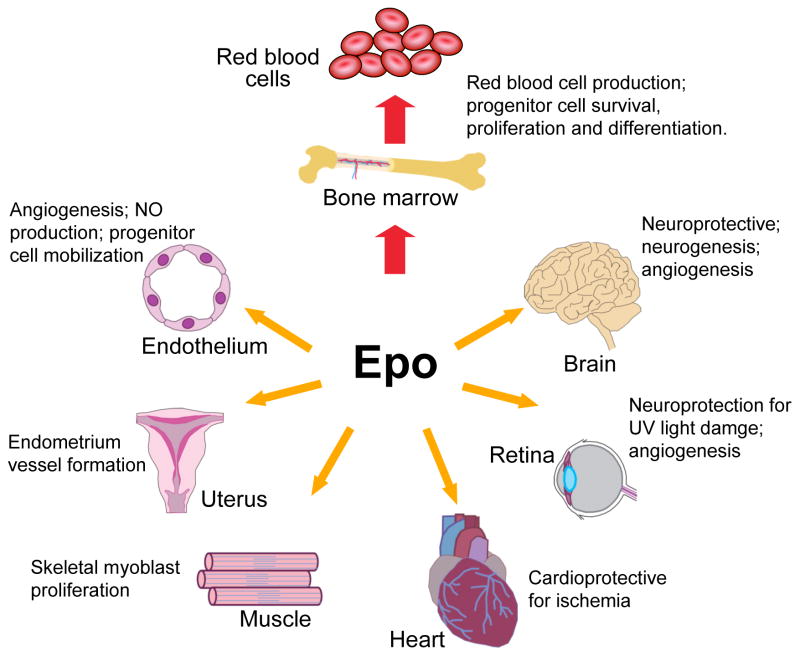
Since the isolation and purification of erythropoietin (EPO) in 1977, the essential role of EPO for mature red blood cell production has been well established. The cloning of the EPO gene and production of recombinant human EPO led to the widespread use of EPO in treating patients with anaemia. However, the biological activity of EPO is not restricted to regulation of erythropoiesis. EPO receptor (EPOR) expression is also found in endothelial, brain, cardiovascular and other tissues, although at levels considerably lower than that of erythroid progenitor cells. This review discusses the survival and proliferative activity of EPO that extends beyond erythroid progenitor cells. Loss of EpoR expression in mouse models provides evidence for the role of endogenous EPO signalling in nonhaematopoietic tissue during development or for tissue maintenance and/or repair. Determining the extent and distribution of receptor expression provides insights into the potential protective activity of EPO in brain, heart and other nonhaematopoietic tissues.


Figures

Figure 1
Figure 1. Hypoxia induction of erythropoietin gene expression
(a) At normoxia, the hypoxia-inducible factor HIF-α is hydroxylated at its prolines by prolyl hydroxylase (PHD), which enables interaction with the von Hippel–Lindau (VHL) protein and ubiquitin (Ub); HIF-α is then polyubiquitinated and degraded by the proteasome. (b) Under hypoxic conditions, HIF-α is stabilised and translocated to the nucleus, where it binds its partner ARNT/HIF-1β to activate erythropoietin (EPO) gene transcription via binding to a 3′ enhancer (Ref. 27). Other factors also contribute to EPO transcription through stabilisation of a macromolecular complex at this 3′ site and reinforcement of enhancer–promoter contact: these include HNF4, p300 (EP300), SMAD3 and SP1 ((Ref. 29, 47, 210) (Ref. 55) Please fix reference format. The open arrow indicates that GATA factors can bind to the 5′ promoter of EPO, and can act as an inducer (GATA4) or repressor (GATA2) in EPO-expressing cells and as a repressor (GATA2/GATA3) in nonexpressing cells (Ref. 16, 55). The dashed arrow indicates that WT1 contributes to tissue-specific EPO expression in hepatocytes (Ref. 46). The five exons of the EPO gene (shaded) and the 3′ transcribed untranslated region (open) are shown schematically on the representation of DNA. Abbreviations: ARNT, arylhydrocarbon receptor nuclear translocator; p300, E1A binding protein p300 (EP300; also known as CBP/p300); HNF4, hepatocyte nuclear factor 4; SMAD3, SMAD family member 3; SP1, Sp1 transcription factor.
Figure 2
Figure 2. Erythropoietin signalling pathways
The binding of erythropoietin (EPO) to its homodimeric receptor (EPOR) results in a conformation change and transphosphorylation of associated tyrosine kinase JAK2. JAK2 activation results in phosphorylation of EPOR and STATs. Activated STATs dimerise and translocate to the nucleus to affect specific downstream gene transcription. JAK2 also activates other signal transduction pathways, including the phosphoinositide 3-kinase (PI3K/AKT), mitogen-activated protein kinase (MAPK), and nuclear factor (NF) κB (p50 and p65) pathways in nonhaematopoietic cells. In EPOR, the four conserved cysteines (thin lines) and the proximal conserved Trp-Ser-X-Trp-Ser motif (thick line) in the extracellular domain, as well as the conserved Box1/Box2 regions (heavy lines) in the membrane-proximal cytoplasmic domain are indicated. Abbreviations: IκB, inhibitor of κB; JAK, Janus kinase; p50 and p65, NF-κB subunits STAT, signal transducer and activator of transcription’.
Figure 3
Figure 3. Embryonic erythropoietin receptor expression beyond erythroid tissues
(a) To visualise erythropoietin receptor (EPOR) expression in nonhaematopoietic tissue, a lacZ reporter gene driven by the EPOR promoter was used to produce transgenic mice. In brain, these mice exhibit expression in the neural tube at embryonic day E10.5 (from (Ref. 131) with permission). (b) At embryonic day 12.5 (left) and 13.5 (right), reporter gene expression resembles developing skeletal muscle (from. (Ref. 60) with permission).
Figure 4
Figure 4. Erythropoietin activity in multiple tissues
Erythropoietin (EPO) acts primarily to regulate erythropoiesis in the bone marrow by stimulating erythroid progenitor cell survival, proliferation and differentiation to produce mature red blood cells. EPO receptor expression on endothelial cells, the endometrium (lining) of the uterus, skeletal muscle myoblasts, the heart, and endothelial cells and neural cells in the retina and brain allows EPO to also act as a survival or mitogenic factor on these nonhaematopoietic cells, providing the potential for a response to EPO in multiple tissues.

References

    1. Funakoshi-Tago M, et al. Jak2 FERM domain interaction with the erythropoietin receptor regulates Jak2 kinase activity. Mol Cell Biol. 2008;28:1792–1801. - PMC - PubMed
    1. Miyake T, Kung CK, Goldwasser E. Purification of human erythropoietin. J Biol Chem. 1977;252:5558–5564. - PubMed
    1. Lin FK, et al. Cloning and expression of the human erythropoietin gene. Proc Natl Acad Sci U S A. 1985;82:7580–7584. - PMC - PubMed
    1. Jacobs K, et al. Isolation and characterization of genomic and cDNA clones of human erythropoietin. Nature. 1985;313:806–810. - PubMed
    1. Livnah O, et al. Crystallographic evidence for preformed dimers of erythropoietin receptor before ligand activation. Science. 1999;283:987–990. - PubMed

Further reading, resources and contacts

    1. Hodges VM, et al. Pathophysiology of anemia and erythorcytosis. Crit Rev Oncol Hematol. 2007;64:139–158. This review describes anaemia and erythrocytosis in relation to erythropoietin (EPO) levels in diagnosis and treatment. - PubMed
    1. Sasaki R. Pleiotropic functions of erythropoietin. Intern Med. 2003;42:142–149. This review highlights the early observations of EPO production in the central nervous system and reproductive organs, including the author’s pioneering work on EPO expression and function in these tissues. - PubMed
    1. Jelkmann W, et al. The erythropoietin receptor in normal and cancer tissues. Crit Rev Oncol/Hematol. 2008;67:39–61. This paper reviews EPO receptor expression and function in tumour tissues and the implications for administration of EPO to treat anaemia in cancer patients. - PubMed
    1. Sytkowski AJ. Erythropoietin: Blood, Brain and Beyond. Wiley-VCH; Weinheim, Germany: 2004. This book provides an overview of the history, developmental biology, transcription regulation and biochemistry of EPO in the context of erythropoiesis and EPO therapy in anaemia in relation to a variety of clinical conditions. Other topics include EPO in nonhaematopoietic tissues and in elite athletics.
    1. Websites:

      This page from the Information Center for Sickle Cell and Thalassemic Disorders website includes an overview of the relationship between iron and erythropoietin activity in red blood cell production: http://sickle.bwh.harvard.edu/iron_epo.html

Publication types

MeSH terms