

Review
doi: 10.1016/j.bbalip.2008.10.009.
Epub 2008 Nov 7.
The life of lipid droplets
Affiliations
- PMID: 19041421
- PMCID: PMC2782899
- DOI: 10.1016/j.bbalip.2008.10.009
Item in Clipboard
Review
The life of lipid droplets
Biochim Biophys Acta.
2009 Jun.
Abstract
Lipid droplets are the least characterized of cellular organelles. Long considered simple lipid storage depots, these dynamic and remarkable organelles have recently been implicated in many biological processes, and we are only now beginning to gain insights into their fascinating lives in cells. Here we examine what we know of the life of lipid droplets. We review emerging data concerning their cellular biology and present our thoughts on some of the most salient questions for investigation.
Figures
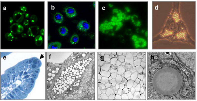
Examples of lipid droplets in eukaryotic cells. Lipid droplets stained with BODIPY (green) in (a) S. cerevisiae, (b) Drosophila S2 cells [nuclei are stained blue with 4′,6-diamidino-2-phenylindole (DAPI)], and (c) murine adipocytes derived from embryonic fibroblasts (image courtesy of R. Streeper). (d) Lipid droplets (bright round organelles) in a single adipocyte derived from OP9 cells (phase contrast, image courtesy of C. Harris). (e) Lipid droplets stained with osmium tetroxide in intestinal enterocytes (arrow indicates an enterocyte that is filled with numerous dark-stained lipid droplets). (f) Electron micrograph of macrophage foam cells in a murine atherosclerotic lesion. Lipid droplets appear as round, empty objects. (g) Section of murine white adipose tissue showing large unilocular lipid droplets (empty spaces) that occupy most of the cytoplasm in white adipocytes. (h) High magnification electron micrograph of a single lipid droplet (large amorphous sphere) in a rat hepatoma cell (image courtesy of S. Stone and J. Wong).
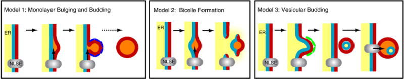
Models of lipid-droplet biogenesis. (Left) Model 1: Lipid droplet biogenesis by ER budding. Neutral lipids (orange) are synthesized by neutral lipid-synthesizing enzymes (NLSE) and bulge from the outer leaflet of the ER membrane (red). The nascent droplet may be coated by proteins (dark blue) that facilitate the budding process. (Middle) Model 2: Bilayer excision. Newly synthesized neutral lipids accumulate between the inner (blue) and outer (red) leaflets of the ER membrane and cause bulging. This entire lipid lens is then excised from the ER, leaving a transient hole in the membrane. ER contents (yellow) might leak through this hole into the cytosol. (Right) Model 3: Vesicular budding. A vesicle containing both leaflets of the ER membrane (red and blue) and a lumen (yellow) is formed by the vesicular budding machinery (green) at the ER membrane. The vesicle is subsequently tethered to the ER, where NLSEs (grey) fill the intramembranous space with neutral lipids (orange). The luminal space (yellow) is compressed, and its contents may leak into the cytosol. This process may trap luminal proteins within a compartment of the lipid droplet.
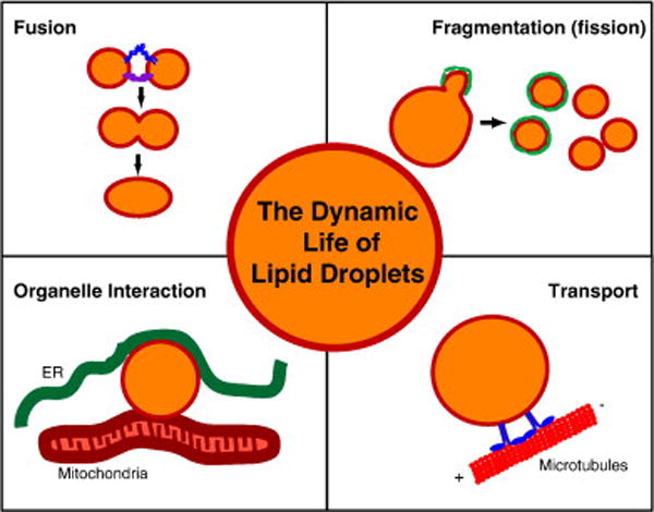
Dynamic processes linked to lipid-droplet biology. Different events in the dynamic life of a lipid droplet (orange interior representing the neutral lipids and a red line indicating the bounding monolayer of phospholipids) are shown in the four panels. In the “fusion” panel, blue and purple protein complexes represent SNARE proteins and tethering complexes, respectively. In the “fragmentation” panel, green proteins covering the lipid droplet during fragmentation indicate COPI/Arf1 coat proteins. During lipid droplet transport, motor proteins (blue) mediate the movement of droplets along microtubules (red).
Similar articles
-
Lipid droplet-organelle interactions; sharing the fats.Biochim Biophys Acta. 2009 Jun;1791(6):441-7. doi: 10.1016/j.bbalip.2008.07.004. Epub 2008 Jul 30. Biochim Biophys Acta. 2009. PMID: 18708159 Review.
-
Lipid droplets as dynamic organelles connecting storage and efflux of lipids.Biochim Biophys Acta. 2009 Jun;1791(6):448-58. doi: 10.1016/j.bbalip.2008.08.001. Epub 2008 Aug 13. Biochim Biophys Acta. 2009. PMID: 18775796 Review.
-
Hepatic stellate cell lipid droplets: a specialized lipid droplet for retinoid storage.Biochim Biophys Acta. 2009 Jun;1791(6):467-73. doi: 10.1016/j.bbalip.2008.11.001. Epub 2008 Nov 24. Biochim Biophys Acta. 2009. PMID: 19071229 Free PMC article. Review.
-
PAT proteins, an ancient family of lipid droplet proteins that regulate cellular lipid stores.Biochim Biophys Acta. 2009 Jun;1791(6):419-40. doi: 10.1016/j.bbalip.2009.04.002. Epub 2009 Apr 16. Biochim Biophys Acta. 2009. PMID: 19375517 Free PMC article. Review.
-
Biogenesis of cytoplasmic lipid droplets: from the lipid ester globule in the membrane to the visible structure.Biochim Biophys Acta. 2009 Jun;1791(6):399-407. doi: 10.1016/j.bbalip.2008.10.002. Epub 2008 Oct 21. Biochim Biophys Acta. 2009. PMID: 18996222 Review.
Cited by
-
Regulation of lipid droplet size in mammary epithelial cells by remodeling of membrane lipid composition-a potential mechanism.PLoS One. 2015 Mar 10;10(3):e0121645. doi: 10.1371/journal.pone.0121645. eCollection 2015. PLoS One. 2015. PMID: 25756421 Free PMC article.
-
Lipid droplets and cellular lipid metabolism.Annu Rev Biochem. 2012;81:687-714. doi: 10.1146/annurev-biochem-061009-102430. Epub 2012 Apr 13. Annu Rev Biochem. 2012. PMID: 22524315 Free PMC article. Review.
-
Energy and Dynamics of Caveolae Trafficking.Front Cell Dev Biol. 2021 Jan 21;8:614472. doi: 10.3389/fcell.2020.614472. eCollection 2020. Front Cell Dev Biol. 2021. PMID: 33692993 Free PMC article. Review.
-
Microsomal Triglyceride Transfer Protein (MTP) Associates with Cytosolic Lipid Droplets in 3T3-L1 Adipocytes.PLoS One. 2015 Aug 12;10(8):e0135598. doi: 10.1371/journal.pone.0135598. eCollection 2015. PLoS One. 2015. PMID: 26267806 Free PMC article.
-
Lipid droplet formation on opposing sides of the endoplasmic reticulum.J Lipid Res. 2012 Sep;53(9):1800-10. doi: 10.1194/jlr.R028290. Epub 2012 Jun 14. J Lipid Res. 2012. PMID: 22701043 Free PMC article. Review.
References
-
- Sessa G, Weissmann G. Phospholipid spherules (liposomes) as a model for biological membranes. J Lipid Res. 1968;9:310–318. - PubMed
-
- Cohen A, Razani B, Schubert W, Williams T, Wang X, Iyengar P, Brasaemle D, Scherer P, Lisanti M. Role of caveolin-1 in the modulation of lipolysis and lipid droplet formation. Diabetes. 2004;53:1261–1270. - PubMed
-
- Vance J. Molecular and cell biology of phosphatidylserine and phosphatidylethanolamine metabolism. Prog Nucleic Acid Res Mol Biol. 2003;75:69–111. - PubMed
-
- Bartz R, Zehmer J, Zhu M, Chen Y, Serrero G, Zhao Y, Liu P. Dynamic activity of lipid droplets: protein phosphorylation and GTP-mediated protein translocation. J Proteome Res. 2007;6:3256–3265. - PubMed
Publication types
MeSH terms
Substances
Grants and funding
LinkOut - more resources
Full Text Sources
Other Literature Sources
