Transcriptional regulation of mouse L-selectin
- PMID: 19041738
- PMCID: PMC2650851
- DOI: 10.1016/j.bbagrm.2008.10.004
Transcriptional regulation of mouse L-selectin
Abstract
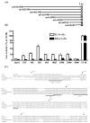
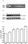
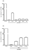
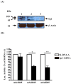
L-selectin mediates the initial tethering and rolling of lymphocytes in high endothelial venules. To study the transcriptional regulation of mouse L-selectin, we cloned 4.5 kb 5'-flanking sequences of the mouse sell. Luciferase analysis of serial 5'-deletion mutants showed that the first 285 bp was sufficient to drive high promoter activity in EL4 cells, but not in Sell-negative HeLa cells, suggesting that this fragment harbors the minimal mouse sell promoter and contains cis-elements for lymphocyte-specific expression. Site-directed mutagenesis and chromatin immunoprecipitation showed that Mzf1, Klf2, Sp1, Ets1, and Irf1 bind to and activate the mouse sell promoter. Over expression of these transcription factors in EL4 cells increased expression of sell mRNA. Silencing the expression of Sp1 by siRNA significantly decreased sell promoter activity in EL4 cells. We conclude that sell transcription is regulated by Mzf1, Klf2, Sp1, Ets1, and Irf1.
Figures


Similar articles
-
A novel functional interaction between the Sp1-like protein KLF13 and SREBP-Sp1 activation complex underlies regulation of low density lipoprotein receptor promoter function.J Biol Chem. 2006 Feb 10;281(6):3040-7. doi: 10.1074/jbc.M509417200. Epub 2005 Nov 22. J Biol Chem. 2006. PMID: 16303770
-
The PP2A-Aβ gene is regulated by multiple transcriptional factors including Ets-1, SP1/SP3, and RXRα/β.Curr Mol Med. 2012 Sep;12(8):982-94. doi: 10.2174/156652412802480916. Curr Mol Med. 2012. PMID: 22827437
-
The IFN-gamma-dependent suppressor of cytokine signaling 1 promoter activity is positively regulated by IFN regulatory factor-1 and Sp1 but repressed by growth factor independence-1b and Krüppel-like factor-4, and it is dysregulated in psoriatic keratinocytes.J Immunol. 2010 Aug 15;185(4):2467-81. doi: 10.4049/jimmunol.1001426. Epub 2010 Jul 19. J Immunol. 2010. PMID: 20644166
-
Transcriptional regulation of the mouse PNRC2 promoter by the nuclear factor Y (NFY) and E2F1.Gene. 2005 Nov 21;361:89-100. doi: 10.1016/j.gene.2005.07.012. Epub 2005 Sep 21. Gene. 2005. PMID: 16181749
-
ETS1 and SP1 drive DHX15 expression in acute lymphoblastic leukaemia.J Cell Mol Med. 2018 May;22(5):2612-2621. doi: 10.1111/jcmm.13525. Epub 2018 Mar 7. J Cell Mol Med. 2018. PMID: 29512921 Free PMC article.
Cited by
-
Foxo: in command of T lymphocyte homeostasis and tolerance.Trends Immunol. 2011 Jan;32(1):26-33. doi: 10.1016/j.it.2010.10.005. Epub 2010 Nov 23. Trends Immunol. 2011. PMID: 21106439 Free PMC article. Review.
-
FOXO1 Up-Regulates Human L-selectin Expression Through Binding to a Consensus FOXO1 Motif.Gene Regul Syst Bio. 2012;6:139-49. doi: 10.4137/GRSB.S10343. Epub 2012 Oct 29. Gene Regul Syst Bio. 2012. PMID: 23133314 Free PMC article.
-
ECM1 controls T(H)2 cell egress from lymph nodes through re-expression of S1P(1).Nat Immunol. 2011 Feb;12(2):178-85. doi: 10.1038/ni.1983. Epub 2011 Jan 9. Nat Immunol. 2011. PMID: 21217760
-
Transiently reduced PI3K/Akt activity drives the development of regulatory function in antigen-stimulated Naïve T-cells.PLoS One. 2013 Jul 11;8(7):e68378. doi: 10.1371/journal.pone.0068378. Print 2013. PLoS One. 2013. PMID: 23874604 Free PMC article.
-
L-selectin: A Major Regulator of Leukocyte Adhesion, Migration and Signaling.Front Immunol. 2019 May 14;10:1068. doi: 10.3389/fimmu.2019.01068. eCollection 2019. Front Immunol. 2019. PMID: 31139190 Free PMC article. Review.
References
-
- Chao CC, Jensen R, Dailey MO. Mechanisms of L-selectin regulation by activated T cells. J Immunol. 1997;159:1686–1694. - PubMed
-
- Ley K, Kansas GS. Selectins in T-cell recruitment to non-lymphoid tissues and sites of inflammation. Nat Rev Immunol. 2004;4:325–335. - PubMed
-
- Rainer TH. L-selectin in health and disease. Resuscitation. 2002;52:127–141. - PubMed
Publication types
MeSH terms
Substances
Grants and funding
LinkOut - more resources
Full Text Sources
Miscellaneous

