. 2008 Nov 28;135(5):865-78.
doi: 10.1016/j.cell.2008.09.045.

Deregulation of scribble promotes mammary tumorigenesis and reveals a role for cell polarity in carcinoma

Affiliations

Deregulation of scribble promotes mammary tumorigenesis and reveals a role for cell polarity in carcinoma

Lixing Zhan et al. Cell. .

Abstract

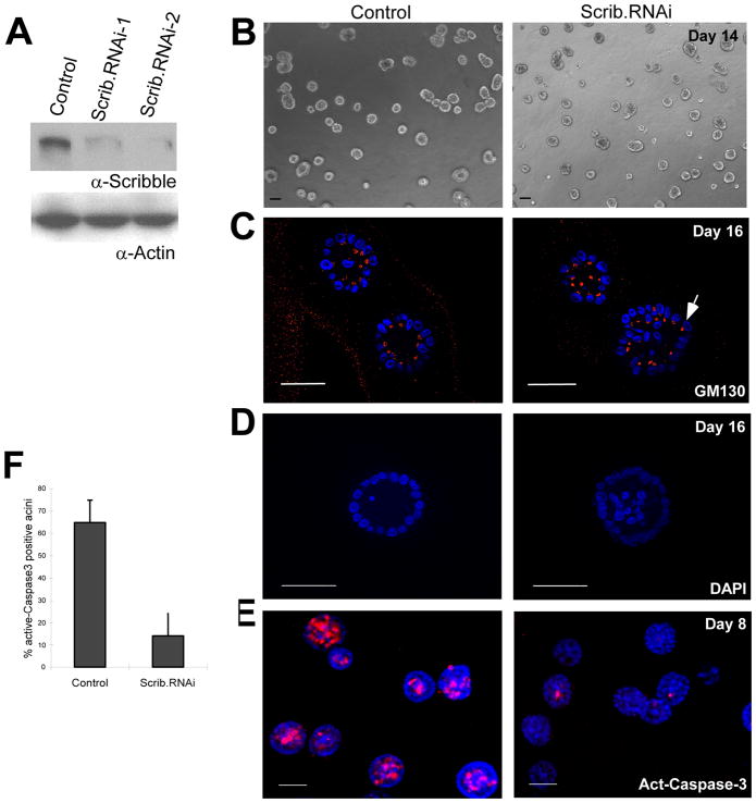
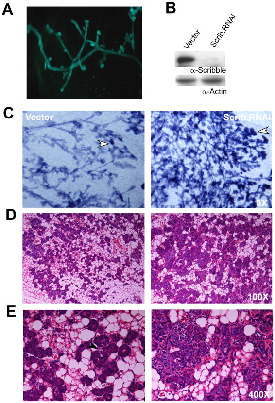
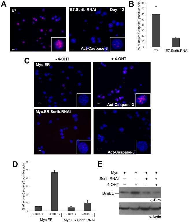
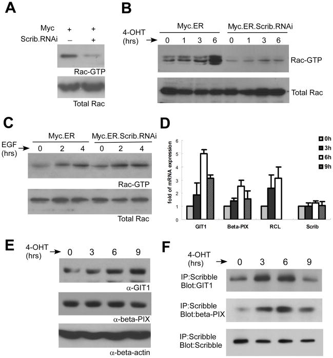
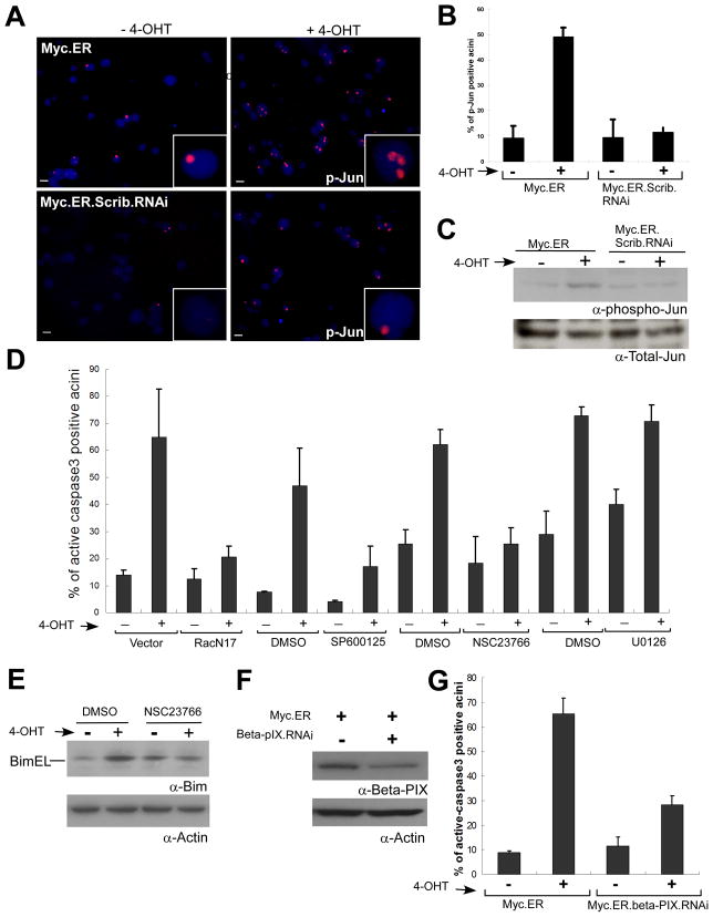
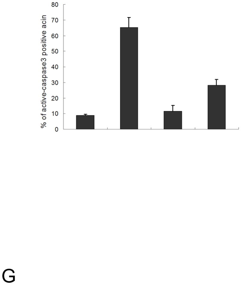
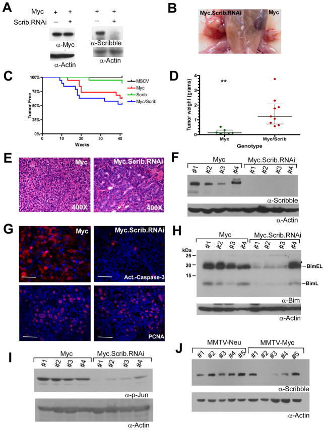
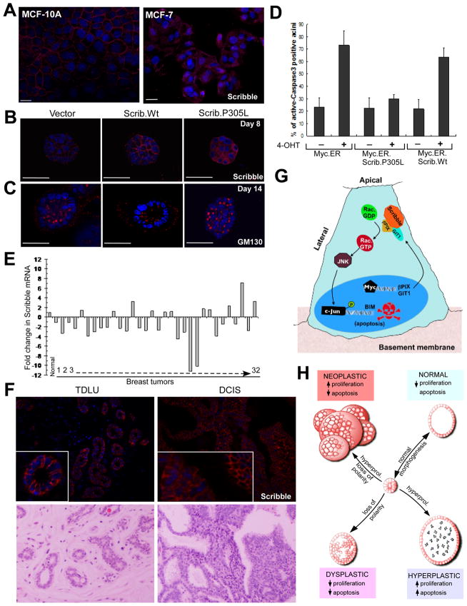
Loss of cell polarity proteins such as Scribble induces neoplasia in Drosophila by promoting uncontrolled proliferation. In mammals, the role that polarity proteins play during tumorigenesis is not well understood. Here, we demonstrate that depletion of Scribble in mammary epithelia disrupts cell polarity, blocks three-dimensional morphogenesis, inhibits apoptosis, and induces dysplasia in vivo that progress to tumors after long latency. Loss of Scribble cooperates with oncogenes such as c-myc to transform epithelial cells and induce tumors in vivo by blocking activation of an apoptosis pathway. Like depletion, mislocalization of Scribble from cell-cell junction was sufficient to promote cell transformation. Interestingly, spontaneous mammary tumors in mice and humans possess both downregulated and mislocalized Scribble. Thus, we demonstrate that scribble inhibits breast cancer formation and that deregulation of polarity pathways promotes dysplastic and neoplastic growth in mammals by disrupting morphogenesis and inhibiting cell death.


Figures

Figure 1
Figure 1. Scribble loss blocks apoptosis and alters 3D organization of epithelial cells
(A) Scribble expression in stable populations of MCF-10A cells expressing control or independant Scribble shRNAs. (B) Phase images of day 14 control or Scrib.RNAi acini. (C) Golgi orientation was visualized in day 16 acini stained with GM130 (Red, Golgi marker) and DAPI. The arrow points to cell where the Golgi is oriented towards the basal surface of the cell, which is rarely seen in control acini. (D) DAPI stained acini to analyze lumen filling. (E) Control or Scrib.RNAi acini (day 8) immunostained for activated caspase-3 (Red, apoptosis marker), and DAPI. (F) Acini positive for activated caspase-3 were counted and the percentage of the total acini are plotted. (n=3, mean ± SD, >150 acini/experiment). Scale bar = 50μM.
Figure 2
Figure 2. Loss of Scribble induces abnormal morphogenesis in transgenic mammary glands
(A) CD-GFP cells were transplanted into epithelium-free mammary fat pads and imaged by fluorescence microscopy 41 weeks post-transplant. (B) Scribble expression in CD cells expressing Scribble.RNAi. (C). Whole mount analysis of 41 week-old mammary outgrowth from GFP or Scrib.RNAi cells. (D) Low and (E) High magnification images of hematoxylin and eosin (H&E) stained sections of CD-GFP or CD-Scrib.RNAi mammary glands.
Figure 3
Figure 3. Scribble loss blocks apoptosis in the context of Myc and E7
(A) Day eight MCF10A acini expressing HPV16 E7 (E7) alone or in combination with Scribble.RNAi immunostained with activated caspase-3 (Red) and DAPI (Blue) (10x) (B) Percentage of activated-caspase-3 positive acini from (A) (n=3, mean ± SD, >150 acini/experiment) (C) Acini from MCF10A cells with empty vector (control) or expressing Myc fused to estrogen receptor (Myc.ER), with and without Scrib.RNAi were stimulated (+) on day 2 with 4-OHT and immunostained on day 6 for activated caspase-3 (Red) and DNA (Blue). Scale bar, 50 μM (D) Percent of activated caspase-3 positive acini positive from (C) (n=3, mean ± SD, >150 acini/experiment). (E) Day 8 acini were stimulated with 4-OHT for 12 hours and lysates analyzed for Bim expression and an actin loading control.
Figure 4
Figure 4. Myc induces activation of Rac by promoting a Scribble-βPIX/GIT1 interaction
(A) Cell lysates from confluent monolayers grown in the absence of 4-OHT were incubated with GST-Pak1 Rac binding domain and bound Rac (Rac-GTP) was monitored by anti-Rac immunoblots. Total Rac levels were monitored in one tenth of the lysate used for the incubation using anti-Rac immunoblots. Cell lysates from confluent monolayers stimulated with 4-OHT (B) or EGF (C) for indicated times were used to monitor changes in Rac.GTP levels as in (A). (D) Cells were stimulated with 4-OHT for indicated times and RNA was isolated. Quantitative PCR was performed using primers against βPIX, RCL, GAPDH (Glyceraldehyde 3-phosphate dehydrogenase), GIT1, Scrib and β-actin. The data was normalized to GAPDH mRNA levels and fold change in mRNA levels of βPIX, GIT1, Scrib and RCL is shown. (n=3, mean±SD) (E) Myc.ER cells were stimulated for indicated periods of time and total cell lysates were analyzed for changes in GIT1 or βPIX protein levels. (F) Scribble immunoprecipitants were analyzed for co-immunoprecipitated βPIX and GIT1.
Figure 5
Figure 5. Myc induces apoptosis by activating a Rac-JNK-Bim pathway
(A) Myc.ER or Myc.ER.Scrib.RNAi acini were stimulated with 4-OHT from day 2 to 6 and immunostained with p-Jun antibody (Red) and with DAPi (Blue). (B) Graph represents quantitation of changes in percentage of p-Jun positive acini (n=3, >100 acini/per experiment; mean± SD). (C) Protein lysates from acini stimulated for three days with 4-OHT analyzed for changes in phospho-Jun and total Jun levels. (D) Percent of activated caspase-3 positive Myc.ER acini (stimulated with 4-OHT from Day 2 to Day 6) expressing RacN17 (Lentivirally transduced) or treated with JNK inhibitor, SP600125 (5μM), Rac inhibitor NSC23766 (50mM) or MEK inhibitor U0126 (1.0μM) (n=3, mean ± SD, >150 acini/experiment). (E) Lysates from Myc.ER acini treated with NSC23766 from day 2 to day 4 analyzed for changes in Bim-EL levels. (F) Myc.ER cell populations infected with shRNA targeting expression of βPIX. (F)Myc.ER or Myc.ER.β.PIX.RNAi acini were stimulated with 4-OHT and percent of acini positive for activated-casapse-3, was determined (n=3, mean ± SD, >150 acini/experiment).
Figure 6
Figure 6. Loss of Scribble cooperates with Myc to induce mammary tumors
(A)Expression of Myc and Scribble in CD.Myc cells alone or in combination with Scrib.RNAi. (B) Representative mammary tumors in #4 and #9 mammary fat pads transplanted with Myc.Scrib.RNAi and Myc cells respectively, 13 weeks post-transplantation. (C) Kaplan-Meier curve for tumor free animals transplanted with control (MSCV, n= 20); Myc (n= 19) Scrib.RNAi (n= 20) or Myc.Scrib.RNAi (n= 19). (D) Myc (n=7) or Myc.Scrib.RNAi tumors (n=9) were weighed at the time of isolation and the distribution plotted. The solid line represents the median value and the inter-quartile range is indicated. The statistical significance was calculated using two-tailed t test (p = 0.003). (E) Tumors were fixed, paraffin embedded, sectioned and stained with H&E. (F) Tumors were lysed and Scribble expression was analyzed. (G) Myc or Myc.Scrib.RNAi tumor tissue were stained by indirect immunoflorescence for activated-caspase-3 (Red) or PCNA (Red) and DAPI (Blue). (n=3 representative images shown). Four tumors were analyzed for changes in Bim expression(H) and phospho-Jun (I) with loading controls. (J) Protein lysates from spontaneous MMTV-Neu and MMTV-Myc driven mammary tumors were analyzed for changes in levels of Scribble protein expression (actin loading control).
Figure 7
Figure 7. Mislocalization of Scribble phenocopies loss of scribble expression
(A) MCF-10A and MCF-7 cells on monolayer cultures were immunostained with Scribble (Red). (B) Day 8 Myc.ER acini (Vector) or cells expressing wild type Scribble (Scrib.Wt) or P305L mutant of Scribble (Scrib.P305L) were immunostained with Scribble antibody (Red) and DAPI (Blue). An optical section through the middle of the acini is shown. (C) Day 14 acini were immunostained with GM130 (Red) and DAPI (Blue) and an optical section through the middle of the acini shown. (D) Percent of acini that stains positive for activated caspase-3 were determined (n=3, mean ± SD, >150 acini/experiment). (E) RNA isolated from 32 primary human breast tumors and four normal breast tissues were analyzed for abundance of Scribble mRNA by quantitative RT-PCR. The graph represents fold change over the average of values obtained from four normal breast samples. (F) Normal human breast or a DCIS tissue section was immunostained using anti-Scribble antibodies (top panel) and a corresponding section was stained with H&E (lower panel). The inserts show higher magnification images to highlight changes in Scribble localization. (G) Summary of the results and a model for how Scribble regulates Myc-induced apoptosis in polarized epithelial cells. (H) A working model for how interaction between cell polarity pathways and oncogenes can be studies with the context of 3D morphogenesis. Remodeling epithelia cycle between proliferation and morphogenesis (indicated by the bi-directional arrow representing the morphogenesis cycle). Hyperplastic growth: achieved by hyperproliferation without loss of polarity (eg., E7) that is coupled to a compensatory increase in apoptosis(grey cells in the lumen). Dysplastic growth: due to disruption of cell polarity and morphogenesis pathways in the presence of normal proliferation cues. Neoplastic growth: due combination of hyperproliferation and loss polarity/morphogenesis achieved due to two cooperating events such as Myc and Scribble loss or due to activation of strong oncogenes such as ErbB2, and RasV12.

Similar articles

Cited by

References

    1. Aguilar-Cordova E, Strange R, Young LJ, Billy HT, Gumerlock PH, Cardiff RD. Viral Ha-ras mediated mammary tumor progression. Oncogene. 1991;6:1601–1607. - PubMed
    1. Allred CA, Hilsenback SG, Mohsin SK. Biological features of human premaligant breast disease. In: Harris J, Lippman ME, Morrow M, Osborne K, editors. Diseases of the Breast. Philadelphia: Lippincott Williams & Wilkins; 2004. pp. 507–520.
    1. Allred DC, Mohsin SK, Fuqua SA. Histological and biological evolution of human premalignant breast disease. Endocr Relat Cancer. 2001;8:47–61. - PubMed
    1. Amundadottir LT, Johnson MD, Merlino G, Smith GH, Dickson RB. Synergistic interaction of transforming growth factor alpha and c-myc in mouse mammary and salivary gland tumorigenesis. Cell Growth Differ. 1995;6:737–748. - PubMed
    1. Amundadottir LT, Nass SJ, Berchem GJ, Johnson MD, Dickson RB. Cooperation of TGF alpha and c-Myc in mouse mammary tumorigenesis: coordinated stimulation of growth and suppression of apoptosis. Oncogene. 1996;13:757–765. - PubMed

Publication types

Substances