. 2008 Dec 4;3(6):625-36.
doi: 10.1016/j.stem.2008.09.018.

Fate tracing reveals the endothelial origin of hematopoietic stem cells

Affiliations

Fate tracing reveals the endothelial origin of hematopoietic stem cells

Ann C Zovein et al. Cell Stem Cell. .

Abstract

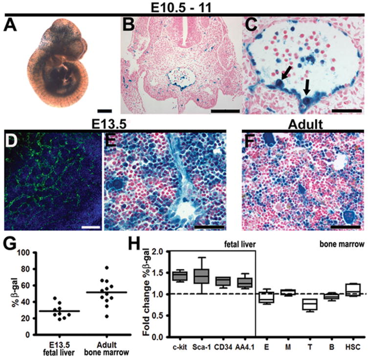
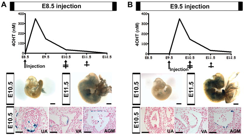
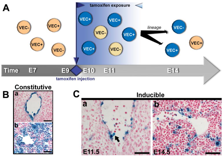
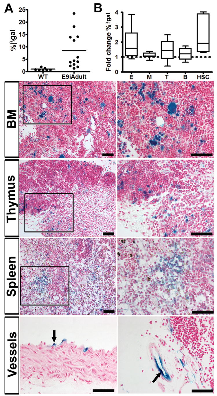
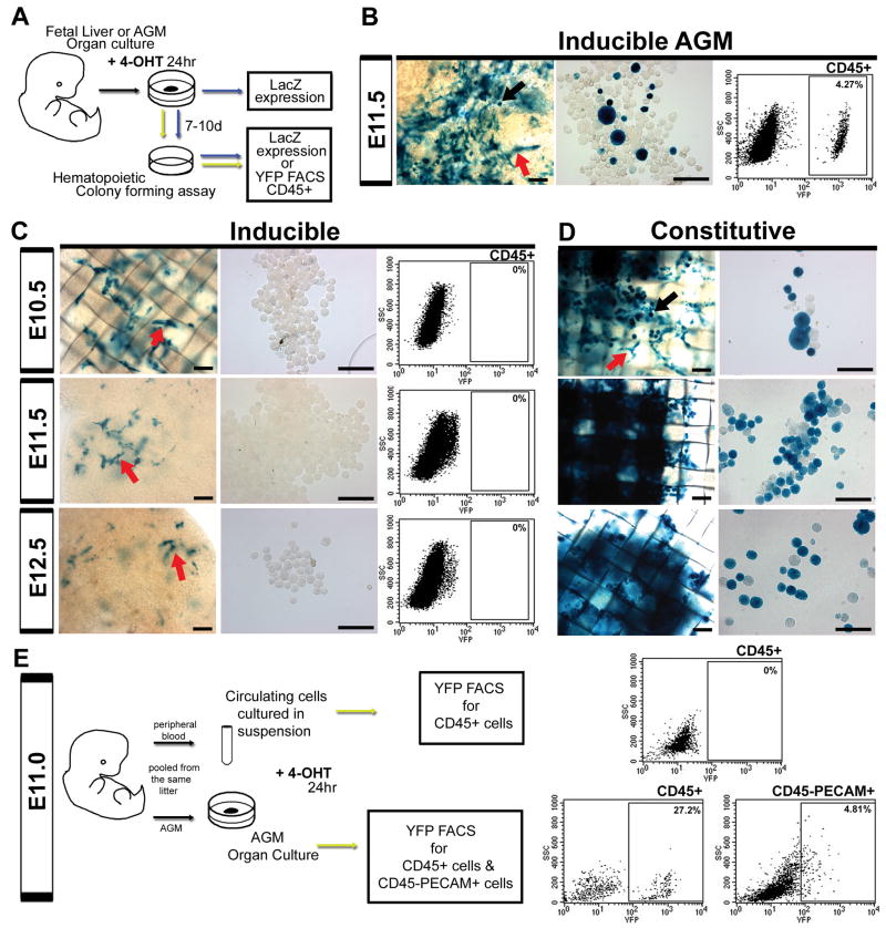
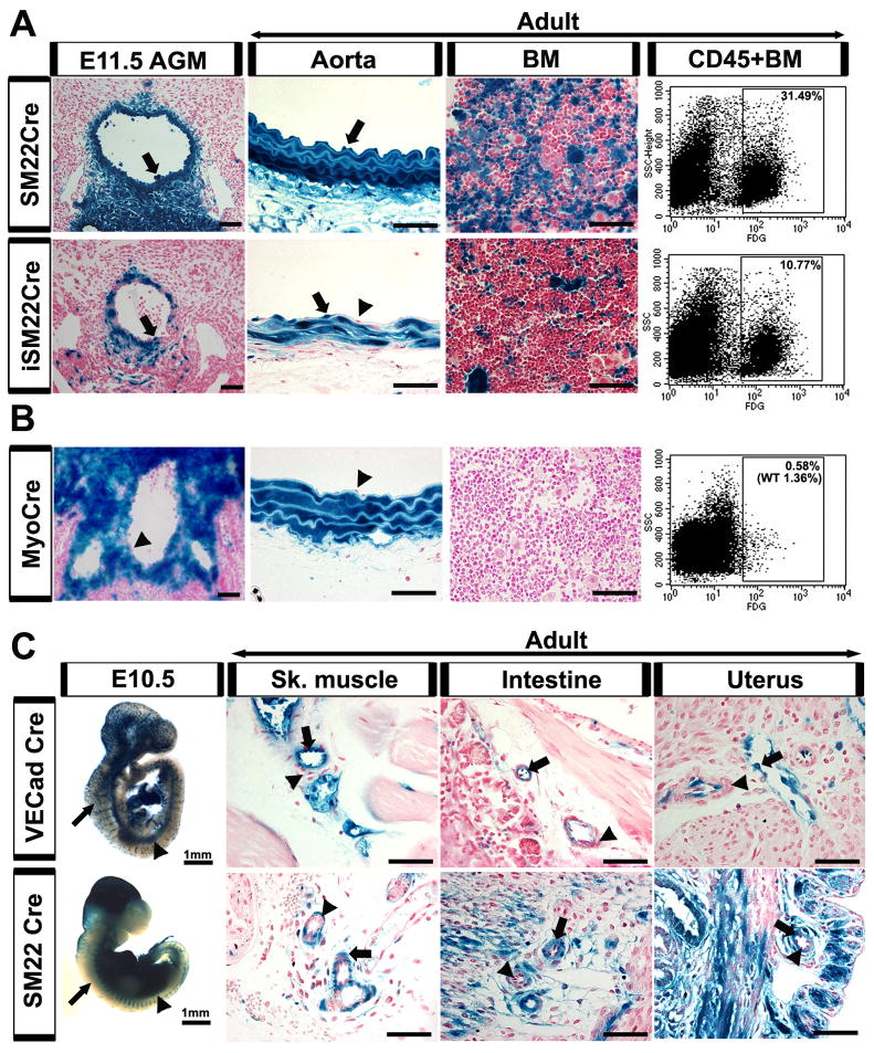
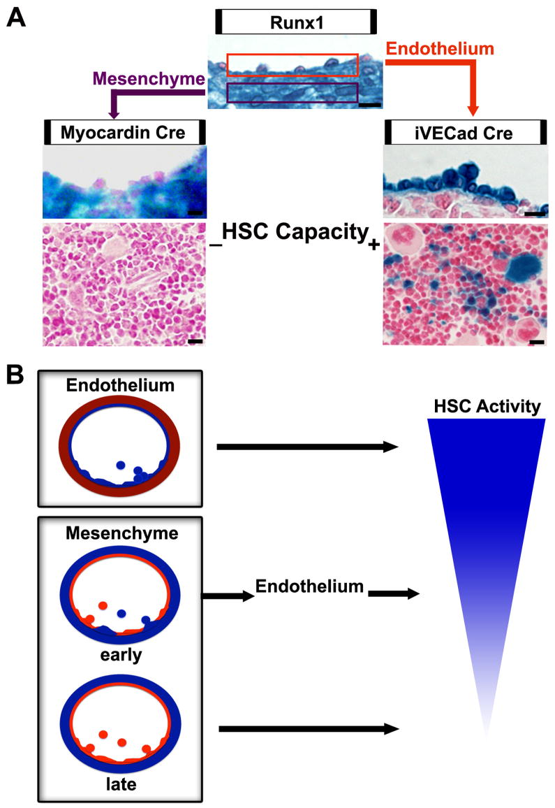
Hematopoietic stem cells (HSCs) originate within the aortic-gonado-mesonephros (AGM) region of the midgestation embryo, but the cell type responsible for their emergence is unknown since critical hematopoietic factors are expressed in both the AGM endothelium and its underlying mesenchyme. Here we employ a temporally restricted genetic tracing strategy to selectively label the endothelium, and separately its underlying mesenchyme, during AGM development. Lineage tracing endothelium, via an inducible VE-cadherin Cre line, reveals that the endothelium is capable of HSC emergence. The endothelial progeny migrate to the fetal liver, and later to the bone marrow, and are capable of expansion, self-renewal, and multilineage hematopoietic differentiation. HSC capacity is exclusively endothelial, as ex vivo analyses demonstrate lack of VE-cadherin Cre induction in circulating and fetal liver hematopoietic populations. Moreover, AGM mesenchyme, as selectively traced via a myocardin Cre line, is incapable of hematopoiesis. Our genetic tracing strategy therefore reveals an endothelial origin of HSCs.


Figures

Figure 1
Figure 1
VE-cadherin Cre labels AGM endothelium and a subset of hematopoietic cells. (A-H) Constitutively active VE-cadherin Cre-recombinase crossed to R26R results in vascular and hematopoietic βgal (blue) labeling. (A) E10.5 Whole mount depicts βgal expression in the vasculature. (B) Histological section of the E11.0 AGM demonstrates labeled endothelium (nuclear counterstain in red). (C) The AGM endothelium and contiguous hematopoietic cells (arrows) exhibit βgal labeling (D) The E13.5 fetal liver demonstrates predominantly vascular VE-cadherin protein expression (green, nuclear stain blue) (E) VE-cadherin βgal labeled progeny consist of both hematopoietic and endothelial cells in the E13.5 fetal liver. (F) Adult bone marrow demonstrates a significant number of labeled hematopoietic cells. (G) Flow cytometry results depict percentage of βgal positive cells within E13.5 fetal liver and adult bone marrow (per animal). (H) Fold change of βgal positive cells per subpopulation compared to total. Fetal liver hematopoietic stem cell populations are enriched within the βgal population (n=5-8, across 3 litters), however this may be due to higher auto-fluorescence levels (Fig. S3). The adult bone marrow lineages: E (erythroid, CD71+/Ter119+), M (myeloid, Gr-1+/Mac-1+), T (T cell, CD3+ or CD4+/CD8+), B (B cell, B220+), HSC (Sca-1+, c-kit+), all demonstrate similar distribution within the βgal compartment (n=6), aside from a slight decrease in T cell lineage βgal expression over total βgal population. Data shown as mean ± sem. Scale bars A = 1mm, B, D = 100μm, C, E, F = 50μm
Figure 2
Figure 2
Differential vascular labeling due to tamoxifen kinetics. (A-B) Top panels demonstrate 4-hydroxytamoxifen levels (4OHT) after maternal injection at either E8.5 (A) or E9.5 (B) based on measured sera levels, see Figure S2. Middle panels depict whole mount embryos, βgal in blue, scale bars = 1mm. Bottom panels demonstrate histological sections of the umbilical artery (UA), vitelline artery (VA), and AGM at E10.5, βgal in blue, nuclear counterstain in red, scale bars = 50μm. (A, middle) Tamoxifen administration at E8.5 results in minimal vascular labeling at E10.5 and E11.5. (A, bottom) Induction at E8.5 results in UA and VA labeling, while minimally labeling the AGM. (B, middle) Induction at E9.5 exhibits less βgal expression at E10.5, but more robust endothelial recombination by E11.5. (B, bottom) E9.5 induction results in labeling of the AGM, but not the UA and VA at E10.5.
Figure 3
Figure 3
Temporal restriction of endothelial tracing by an inducible VE-cadherin Cre system. (A) Schema depicting the inducible Cre/R26R lineage tracing system, where Cre remains inactive until tamoxifen (1mg) is administered at E9.5, resulting in subsequent recombination and labeling during the defined tamoxifen window (12 - 72 hrs). (B) Histological sections depict constitutive Cre/R26R βgal labeling of the AGM endothelium at E10.5 (a), and traced fetal liver cells at E14.5 (b). (Ca) Identification of βgal labeled AGM endothelium and “budding” hematopoietic progeny (arrow) at E11.5, after tamoxifen injection. (Cb) This inducible system traces a substantial historical population that can be found in the fetal liver at E14.5. (B-C) Scale bars = 50μm. βgal activity in blue, nuclear counterstain in red.
Figure 4
Figure 4
VE-cadherin Cre induction at E9.5 results in long-term adult hematopoiesis. (A) Flow cytometry analysis depicts βgal labeled cells in adult bone marrow per animal after embryonic induction, compare to wildtype (WT). (B) Fold change of βgal positive cells per lineage as compared to total (averaged, n=12). All adult bone marrow lineages are labeled: E (erythroid, CD71+/Ter119+), M (myeloid, Gr-1+/Mac-1+), T (T cell, CD3+ or CD4+/CD8+), B (B cell, B220+), HSC (Sca-1+,c-kit+). This is also seen in a cohort (n=6) one year after induction (data averaged into total pool of 12). Data shown as mean ± sem. (Panels) Boxed areas in the left columns are magnified on right. Histological sections depict βgal labeling of hematopoietic cells in the adult bone marrow, thymus, and spleen (n=4). (Bottom) Labeled endothelial cells of the aorta (left) and thymus (right) are traced in the adult after embryonic induction. Scale bars =50um. βgal activity in blue, nuclear counterstain in red.
Figure 5
Figure 5
VE-cadherin Cre fetal liver and peripheral blood hematopoietic populations are endothelial derived. (A) Organ cultures of fetal liver (or AGM) underwent 24 hour 4-hydroxytamoxifen (4OHT) in vitro induction using separate LacZ and EYFP Cre reporter lines. A subset of cultured organs (livers and AGMs) were dissociated and cultured in methylcellulose for 7-10 days, and both organ cultures and cells obtained from hematopoietic assays were evaluated for βgal labeling. Alternatively, cells from hematopoietic assays of the EYFP line underwent FACS analysis of EYFP expression after gating for CD45 expression. (B-D) Organ cultures are shown in left panels (scale bars = 100μm) and cells removed from hematopoietic assays in middle panels (scale bars = 50μm), red arrows indicate endothelial morphology, black arrows hematopoietic morphology, βgal expression in blue. (B-C, right panels) Gated EYFP expression within the CD45+ compartment. (B) Induction of AGM at E11.5 resulted in both endothelial and hematopoietic βgal activity (arrows) quantified as 4.27% EYFP labeled CD45+ cells. (C) When fetal liver is induced for 24 hrs at E10.5, E11.5, and E12.5, βgal labeling is restricted to apparent endothelial populations that do not produce hematopoietic cells in culture. (D) In contrast, the constitutive system demonstrates βgal labeling in both hematopoietic and endothelial populations throughout all time points. (E) Peripheral blood was pooled at E11.0 and induced in vitro for 24 hrs in suspension. Cells were then analyzed for EYFP expression within the CD45+ population. Alternatively, the corresponding AGMs were induced for 24 hrs in organ culture, pooled, and analyzed for EYFP expression within hematopoietic (CD45+) and endothelial (PECAM+ CD45-) compartments. Only the AGM, with endothelial induction, was capable of producing labeled hematopoietic cells.
Figure 6
Figure 6
Subaortic mesenchyme does not contribute to hematopoiesis, except through an endothelial intermediate. (A-C) Labeled cells depicted by arrows, unlabeled by arrowheads. (A, top panel) SM22α Cre crossed with the R26R LacZ reporter line demonstrates labeling not only in the subaortic mesenchyme of the AGM (E11.5), but also within the endothelial layer. The endothelial labeling persists in the adult aorta and results in labeled hematopoietic cells in the adult bone marrow by histology and FACS of βgal expression (31.49% of CD45+ population). (A, bottom panel) To restrict SM22α Cre labeling to a period during AGM hematopoiesis, a tamoxifen inducible SM22α Cre was employed and induced at E9.5. While the mesenchymal contribution to the endothelium was decreased in the AGM (E11.5), there remained a population of labeled endothelium and subsequent hematopoietic cells (10.77%) in the adult. (B) Myocardin Cre crossed to a R26R LacZ reporter line demonstrates labeling of the AGM mesenchyme at E11.5, without contribution to the AGM or adult aortic endothelium, and absence of any hematopoietic contribution to adult bone marrow (0.58% with 1.36% WT background in FACS-gal assay). (C) Whole mount SM22α Cre and VE-cadherin Cre embryos at E10.5 demonstrate similar βgal staining pattern in the dorsal aorta (arrowheads) but not in the intersomitic or surface vessels (arrows). When the two Cre lines are compared in adult vascular beds, the smooth muscle layers are labeled in the SM22α line (arrows) but not in the VE-cadherin line (arrowheads). In contrast, the endothelial layer is labeled in the VE-cadherin Cre (arrows) but not the SM22α Cre (arrowheads). (A-C) βgal activity depicted in blue, nuclear counterstain in red. Scale bars = 50μm, unless otherwise specified.
Figure 7
Figure 7
Midgestation hematopoietic stem cells are endothelial derived. (A) Runx1, a transcription factor critical for definitive hematopoiesis, is expressed in both the AGM endothelium and mesenchyme as exhibited by the Runx1-LacZ transgenic mouse line. When AGM endothelium is selectively labeled using the inducible VE-cadherin Cre crossed to a R26R LacZ reporter line, hematopoietic stem cells are traced to adult bone marrow. However, when the AGM subaortic mesenchyme is selectively labeled, hematopoietic cells are not labeled in the adult bone marrow. Scale bars = 10μm. (B) Endothelium maintains hematopoietic capacity in the AGM. The early transient mesenchymal population of the AGM contributes to the endothelium and subsequently to hematopoiesis. Later AGM mesenchymal populations do not have endothelial potential and therefore do not have hematopoietic capacity.

References

    1. Alva JA, Zovein AC, Monvoisin A, Murphy T, Salazar A, Harvey NL, Carmeliet P, Iruela-Arispe ML. VE-Cadherin-Cre-recombinase transgenic mouse: a tool for lineage analysis and gene deletion in endothelial cells. Dev Dyn. 2006;235(3):759–767. - PubMed
    1. Alvarez-Silva M, Belo-Diabangouaya P, Salaun J, Dieterlen-Lievre F. Mouse placenta is a major hematopoietic organ. Development (Cambridge, England) 2003;130(22):5437–5444. - PubMed
    1. Bertrand JY, Giroux S, Golub R, Klaine M, Jalil A, Boucontet L, Godin I, Cumano A. Characterization of purified intraembryonic hematopoietic stem cells as a tool to define their site of origin. Proceedings of the National Academy of Sciences of the United States of America. 2005;102(1):134–139. - PMC - PubMed
    1. Bollerot K, Pouget C, Jaffredo T. The embryonic origins of hematopoietic stem cells: a tale of hemangioblast and hemogenic endothelium. Apmis. 2005;113(1112):790–803. - PubMed
    1. Breier G, Breviario F, Caveda L, Berthier R, Schnurch H, Gotsch U, Vestweber D, Risau W, Dejana E. Molecular cloning and expression of murine vascular endothelial-cadherin in early stage development of cardiovascular system. Blood. 1996;87(2):630–641. - PubMed

Publication types

MeSH terms