In vitro analog of human bone marrow from 3D scaffolds with biomimetic inverted colloidal crystal geometry
- PMID: 19042018
- PMCID: PMC2650812
- DOI: 10.1016/j.biomaterials.2008.10.041
In vitro analog of human bone marrow from 3D scaffolds with biomimetic inverted colloidal crystal geometry
Abstract
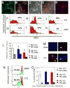
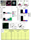
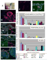
In vitro replicas of bone marrow can potentially provide a continuous source of blood cells for transplantation and serve as a laboratory model to examine human immune system dysfunctions and drug toxicology. Here we report the development of an in vitro artificial bone marrow based on a 3D scaffold with inverted colloidal crystal (ICC) geometry mimicking the structural topology of actual bone marrow matrix. To facilitate adhesion of cells, scaffolds were coated with a layer of transparent nanocomposite. After seeding with hematopoietic stem cells (HSCs), ICC scaffolds were capable of supporting expansion of CD34+ HSCs with B-lymphocyte differentiation. Three-dimensional organization was shown to be critical for production of B cells and antigen-specific antibodies. Functionality of bone marrow constructs was confirmed by implantation of matrices containing human CD34+ cells onto the backs of severe combined immunodeficiency (SCID) mice with subsequent generation of human immune cells.
Conflict of interest statement
The authors declare no conflict of interest.
Figures


Similar articles
-
Cord blood-hematopoietic stem cell expansion in 3D fibrin scaffolds with stromal support.Biomaterials. 2012 Oct;33(29):6987-97. doi: 10.1016/j.biomaterials.2012.06.029. Epub 2012 Jul 15. Biomaterials. 2012. PMID: 22800538
-
Fabrication of Bioactive Inverted Colloidal Crystal Scaffolds Using Expanded Polystyrene Beads.Tissue Eng Part C Methods. 2020 Mar;26(3):143-155. doi: 10.1089/ten.TEC.2019.0333. Epub 2020 Mar 6. Tissue Eng Part C Methods. 2020. PMID: 32031058 Free PMC article.
-
Co-cultured hBMSCs and HUVECs on human bio-derived bone scaffolds provide support for the long-term ex vivo culture of HSC/HPCs.J Biomed Mater Res A. 2016 May;104(5):1221-30. doi: 10.1002/jbm.a.35656. Epub 2016 Feb 12. J Biomed Mater Res A. 2016. PMID: 26779960
-
Biomimetic Materials and Fabrication Approaches for Bone Tissue Engineering.Adv Healthc Mater. 2017 Dec;6(23). doi: 10.1002/adhm.201700612. Epub 2017 Nov 24. Adv Healthc Mater. 2017. PMID: 29171714 Review.
-
An overview of inverted colloidal crystal systems for tissue engineering.Tissue Eng Part B Rev. 2014 Oct;20(5):437-54. doi: 10.1089/ten.TEB.2013.0402. Epub 2014 Apr 24. Tissue Eng Part B Rev. 2014. PMID: 24328724 Review.
Cited by
-
Mesenchymal Stem Cells as a Promising Cell Source for Integration in Novel In Vitro Models.Biomolecules. 2020 Sep 10;10(9):1306. doi: 10.3390/biom10091306. Biomolecules. 2020. PMID: 32927777 Free PMC article. Review.
-
Advancing Tissue Engineering: A Tale of Nano-, Micro-, and Macroscale Integration.Small. 2016 Apr 27;12(16):2130-45. doi: 10.1002/smll.201501798. Epub 2015 Dec 3. Small. 2016. PMID: 27101419 Free PMC article. Review.
-
Multiscale engineering of immune cells and lymphoid organs.Nat Rev Mater. 2019 Jun;4(6):355-378. doi: 10.1038/s41578-019-0100-9. Epub 2019 Apr 3. Nat Rev Mater. 2019. PMID: 31903226 Free PMC article.
-
Maintenance and expansion of hematopoietic stem/progenitor cells in biomimetic osteoblast niche.Cytotechnology. 2010 Oct;62(5):439-48. doi: 10.1007/s10616-010-9297-6. Epub 2010 Sep 10. Cytotechnology. 2010. PMID: 20830608 Free PMC article.
-
Bioengineered implantable scaffolds as a tool to study stromal-derived factors in metastatic cancer models.Cancer Res. 2014 Dec 15;74(24):7229-38. doi: 10.1158/0008-5472.CAN-14-1809. Epub 2014 Oct 22. Cancer Res. 2014. PMID: 25339351 Free PMC article.
References
-
- Wilson A, Trumpp A. Bone-marrow haematopoietic-stem-cell niches. Nat Rev Immunol. 2006;6(2):93–106. - PubMed
-
- Kobari L, Pflumio F, Giarratana MC, Li X, Titeux M, Izac B, et al. In vitro and in vivo evidence for the long-term multilineage (myeloid, B, NK, and T) reconstitution capacity of ex vivo expanded human CD34+ cord blood cells. Exp Hematol. 2000;28:1470–1480. - PubMed
-
- Barker J, Verfaillie C. A novel in vitro model of early human adult B lymphopoiesis that allows proliferation of pro-B cells and differentiation to mature B lymphocytes. Leukemia. 2000;14:1614–1620. - PubMed
-
- Chen J, Brandt JS, Ellison FM, Calado RT, Young NS. Defective stromal cell function in a mouse model of infusion-induced bone marrow failure. Exp Hematol. 2005;33(8):901–908. - PubMed
-
- Punzel M, Moore K, Lemischka I, Verfaillie C. The type of stromal feeder used in limiting dilution assays influences frequency and maintenance assessment of human long-term culture initiating cells. Leukemia. 1999;13:92–97. - PubMed
Publication types
MeSH terms
Substances
Grants and funding
LinkOut - more resources
Full Text Sources
Other Literature Sources

