. 2009 Jan;11(1):56-64.
doi: 10.1038/ncb1812. Epub 2008 Nov 30.

MG53 nucleates assembly of cell membrane repair machinery

Affiliations

MG53 nucleates assembly of cell membrane repair machinery

Chuanxi Cai et al. Nat Cell Biol. 2009 Jan.

Abstract

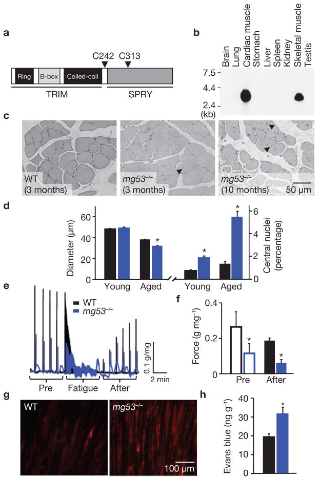
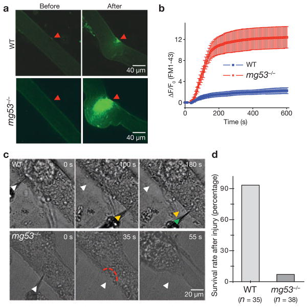
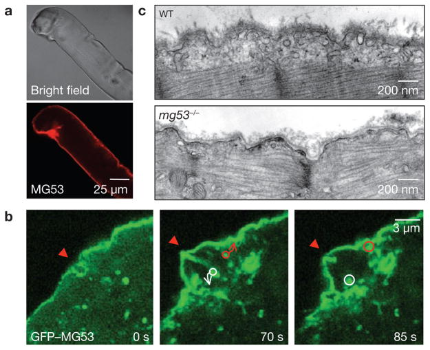
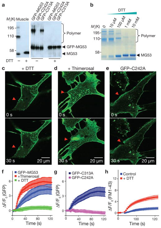
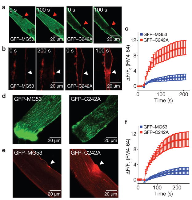
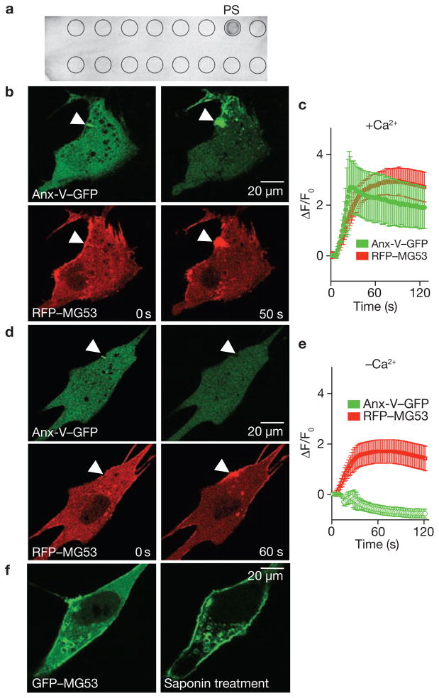
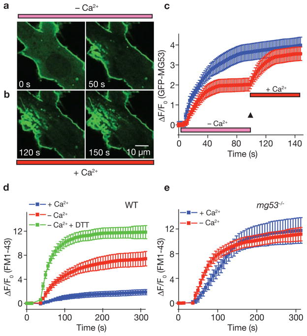
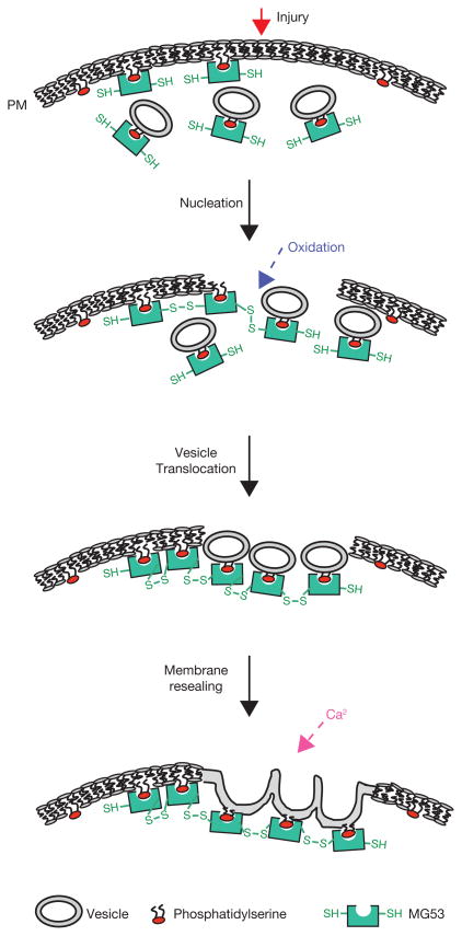
Dynamic membrane repair and remodelling is an elemental process that maintains cell integrity and mediates efficient cellular function. Here we report that MG53, a muscle-specific tripartite motif family protein (TRIM72), is a component of the sarcolemmal membrane-repair machinery. MG53 interacts with phosphatidylserine to associate with intracellular vesicles that traffic to and fuse with sarcolemmal membranes. Mice null for MG53 show progressive myopathy and reduced exercise capability, associated with defective membrane-repair capacity. Injury of the sarcolemmal membrane leads to entry of the extracellular oxidative environment and MG53 oligomerization, resulting in recruitment of MG53-containing vesicles to the injury site. After vesicle translocation, entry of extracellular Ca(2+) facilitates vesicle fusion to reseal the membrane. Our data indicate that intracellular vesicle translocation and Ca(2+)-dependent membrane fusion are distinct steps involved in the repair of membrane damage and that MG53 may initiate the assembly of the membrane repair machinery in an oxidation-dependent manner.


Conflict of interest statement

COMPETING FINANCIAL INTERESTS

The authors declare no competing financial interests.

Figures

Figure 1
Figure 1
Mice null for MG53, a muscle specific TRIM family protein, show progressive muscle pathology. (a) Motif structure of MG53. For detailed alignment of mouse, rabbit and human protein sequences, see Supplementary Information, Fig. S1. (b) Northern blot analysis of mouse tissues shows the specific expression of MG53 in skeletal and cardiac muscles. Total RNA (15 μg per lane) from different mouse tissues was analysed using a cDNA probe following a procedure described previously. (c) Haematoxylin and eosin (H&E) staining of EDL muscle sections illustrates the increased number of central nuclei (arrows) in ageing mg53−/− muscle (10 months), compared with young (3 months) wild-type (WT) or mg53−/− mice. (d) The diameter of muscle fibres in aged (10–11 months) mg53−/− mice (32.1 ± 0.3 μm, n = 541, *P < 0.05 by ANOVA) was decreased, compared with aged (10–11 months) WT controls (37.9 ± 0.5 μm, n = 562), whereas there was no difference in young (3–5 months) WT (48.4 ± 0.5 μm, n = 765) versus mg53−/− (49.5 ± 0.5 μm, n = 673) muscle. Percentage of muscle fibres that with central nuclei in mg53−/− skeletal muscle (5.47 ± 0.01%, *P < 0.05, ANOVA) increased with age when compared with WT (1.25 ± 0.00%). Data are mean ± s.e.m. (e) Trace recordings of contractile performance of intact soleus muscle obtained from ageing mice subjected to 30 min downhill running was assessed using an in vitro voltage stimulation protocol as described previously. The black trace represents WT muscle, blue trace corresponds to mg53−/− muscle. (f) Before fatigue stimulation, the maximal tetanic force, normalized in g mg−1 total protein, was significantly lower in mg53−/− muscle (0.12 ± 0.05), compared with WT (0.27 ± 0.08, n = 4). At 6 min after fatigue stimulation, the WT muscle (0.18 ± 0.02) recovered significantly more than mg53−/− muscle (0.06 ± 0.02, *P < 0.05, ANOVA). (g) Extensive Evans blue staining reveals severe damage in mg53−/− tibialis anterior muscle subjected to a single round of downhill running, compared with minimal staining in WT muscles. (h) Elevated levels of Evans blue dye could be extracted from the gastrocnemius muscle of mg53−/− mice after a single round of eccentric exercise. Data represent mean ± s.d. (n = 4), *P < 0.01.
Figure 2
Figure 2
Defective membrane repair capacity in mg53−/− muscle. (a) Isolated FDB fibres from ageing WT mice can effectively exclude FM1-43 dye after UV-laser induced damage, whereas fibres from ageing mg53−/− mice cannot prevent dye entry. The arrow indicates a wounding site at 401 s after laser injury. Local contraction due to the entry of Ca2+o was greater in mg53−/− muscle, and exceeds that reported previously. (b) Time-dependent accumulation of FM1-43 inside FDB muscle fibre induced by laser damage of the sarcolemmal membrane with Ca2+ present in the extracellular solution. Data are mean ± s.e.m of 30 fibres obtained from WT mice and 18 fibres from mg53−/− mice. (c) Representative images of differentiated myotubes derived from WT or mg53−/− neonates indicate that WT myotubes do not contract after mechanical damage by multiple penetrations with a microelectrode (arrows), whereas mg53−/− myotubes contract after a single injury, reflecting their defective membrane-repair capacity. (d) As these microelectrode penetration experiments were conducted with Ca2+ present in the extracellular solution, the lack of membrane resealing will lead to excessive Ca2+ entry and contraction of myotubes. If the cell did not contract over a 5 min period, it was considered to have survived microelectrode injury. The summary data presented indicates that mg53−/− muscle has markedly compromised ability to reseal cellular membranes following injury.
Figure 3
Figure 3
MG53 facilitates repair of acute membrane damage in muscle cells. (a) Immunostaining of MG53 in isolated WT FDB fibres to illustrate the concentration of MG53 at the injury site. These are representative images from more than 20 different muscle fibres that show damage during isolation. (b) GFP–MG53 expressed in mg53−/− myotubes localizes to intracellular vesicles and sarcolemmal membrane (left). Penetration with a microelectrode leads to accumulation of GFP–MG53 at the injury sites (arrows) at 70 s after injury (right, n = 18). Circles surround individual vesicles containing GFP–MG53 and arrows indicate the path to fusion with the injury site. See Supplementary Information, Movie 7 for visualization of the dynamic process of GFP–MG53 vesicle translocation. (c) Representative electron micrographs of EDL muscle following downhill running exercise. Sub-sarcolemmal accumulation of vesicles was observed in 70 out of 79 muscle fibres examined in WT preparations from two different 6-month-old mice. In contrast, mg53−/− muscle fibres lack accumulation of intracellular vesicles, as 41 out of 104 muscle fibres from two aged-matched mg53−/− animals do not show such accumulation.
Figure 4
Figure 4
Oxidation-mediated MG53 oligomerization serves as a nucleation mechanism for acute membrane repair. (a) Western blot of mouse skeletal muscle (0.2 μg) or C2C12 cell lysates (0.5 μg per lane) from cells transfected with GFP–MG53, GFP–C242A or GFP–C313A. Native MG53 dimers and tetramers from skeletal muscle were observed under oxidizing condition (–DTT), and after addition of 10 mM DTT (+DTT) caused MG53 to de-oligomerize into the monomeric form (left). The GFP–C242A mutant did not form oligomers, even in the oxidized state, whereas GFP–C313A behaved similarly to WT GFP–MG53 (right; n = 9 for skeletal muscle preparation and n = 8 for C2C12 cells). (b) Purified recombinant MG53 protein shows concentration-dependent de-oligomerization in response to DTT. (c) The presence of DTT (5 mM) in the bathing solution of C2C12 cells abolished GFP–MG53 accumulation at injury sites. (d) Addition of thimerosal (2 μM) to the extracellular solution accelerated accumulation of GFP–MG53 at the damage site of a C2C12 cell. (e) GFP–C242A localized to intracellular vesicles and plasma membrane, but did not accumulate at the microelectrode injury sites. Arrowheads indicate the location of microelectrode penetration (ce). (f) Time-course of GFP–MG53 accumulation at injury sites following microelectrode penetration in untreated C2C12 cells and in the presence of DTT or thimerosal. Data represent mean ± s.e.m. (n = 12). (g) Time-course of GFP–C242A and GFP–C313A accumulation at injury sites after microelectrode penetration in C2C12 cells. Data represent mean ± s.e.m. (n = 15). (h) Time-course of FM1-43 entry into WT skeletal muscle fibres in the presence or absence of DTT (10 mM) following laser induced injury. Data represent mean ± s.e.m. (n = 8).
Figure 5
Figure 5
Repair patch formation by MG53 restores cell integrity following acute injury. (a) GFP–MG53 expressed in mg53−/− myotubes translocates to cell injury site after UV-bleaching (left), whereas GFP–C242A remains immobile after photobleaching (right). (b) mg53−/− myotubes transfected with GFP–MG53 (left) or GFP–C242A (right) were assayed for FM4-64 dye entry after UV-laser damage. Arrows indicate the site of laser damage. (c) GFP–MG53 expression (blue) can prevent FM4-64 dye entry, whereas GFP–C242A cannot (red). Data represent mean ± s.e.m. (n = 9). (d) FDB muscle fibres from WT mice were transfected by in vivo electroporation to allow for transient expression of GFP–MG53 (left) and GFP–C242A (right) (see also Supplementary Information, Fig. S5). (e) FDB muscle fibres transfected with GFP–C242A show excessive FM4-64 dye entry following UV-laser wounding (right), compared with those transfected with GFP–MG53 (left; n = 15). (f) Summary data for panel e. Data represent mean ± s.e.m., n = 8.
Figure 6
Figure 6
MG53 binds to phosphatidylserine (PS) to mediate Ca2+-independent vesicle translocation to the injury site. (a ) PIP2-Strip lipid dot blot analysis reveals recombinant MG53 (1 μg ml−1) binds PS but not other membrane lipids, including sphingosine-1-P, phosphatidic acid, phosphotidylcholine, phosphatidylethanolamine and various phosphainositol metabolites. (b) Microelectrode penetration of C2C12 cells co-expressing annexin-V–GFP (top) and RFP–MG53 (bottom) in the presence of 2 mM Ca2+o results in colocalization of annexin-V and MG53 at the injury site. (c) Time-course of annexin-V–GFP and RFP–MG53 accumulation at injury sites following microelectrode penetration into C2C12 cells. RFP–MG53 continued to accumulate at the injury site, whereas annexin-V–GFP accumulation seemed to be biphasic. Membrane resealing probably reduces Ca2+ entry-dependent binding of annexin to PS-enriched membrane surfaces at later time points. Data represent mean ± s.e.m. (n = 16). (d) Removal of Ca2+o prevents movement of annexin-V–GFP (top) to the injury site (arrow), whereas RFP–MG53 can still translocate following membrane disruption (bottom). (e) Time-dependent changes for accumulation of RGP–MG53 and annexin-V–GFP following acute injury of C2C12 cells in the absence of Ca2+o plus 0.5 mM EGTA. Data represent mean ± s.e.m., n = 12. (f) C2C12 myoblasts transfected with GFP–MG53 show translocation of GFP–MG53 to the plasma membrane following treatment with 0.005% saponin in an extracellular solution containing 0 Ca2+ plus 0.5 mM EGTA.
Figure 7
Figure 7
Relative contribution of extracellular Ca2+ and oxidation to membrane repair in skeletal muscle. (a) Injury of GFP–MG53 expressing mg53−/− myotube by penetration with a microelectrode leads to accumulation of GFP–MG53 at the damage sites in an extracellular solution containing 0 Ca2+ and 0.5 mM EGTA (–Ca2+). (b) Rapid perfusion of solution containing 2 mM Ca2+ (+Ca2+) elevated the accumulation and fusion of GFP–MG53 containing vesicles at the injury site. (c) Time-dependent accumulation of GFP–MG53 at the injury sites show a two-step translocation of GFP–MG53 in response to acute damage to the plasma membrane in the absence of Ca2+, followed by addition of 2 mM Ca2+ (red), compared with continuous incubation with 2 mM Ca2+ for mg53−/− myotubes transfected with GFP–MG53 (blue). Data are mean ± s.e.m (n = 12). (d) Time-dependent accumulation of FM1-43 dye inside WT muscle fibre induced by a laser damage of the sarcolemmal membrane with the presence of 2 mM Ca2+o, or absence of Ca2+o (0 Ca2+ plus 0.5 mM EGTA), or incubated with 8 mM DTT and 0.5 mM EGTA in the extracellular solution. Data represent mean ± s.e.m. (n = 12 fibres for each group). (e) Time-dependent accumulation of FM1-43 inside mg53−/− muscle fibre induced by laser damage of the sarcolemmal membrane with the presence of 2 mM Ca2+o, or absence of Ca2+o. Data represent mean ± s.e.m. (n = 12 fibres for each group).
Figure 8
Figure 8
A schematic representation of the proposed function of MG53 in muscle membrane repair. Through interaction with phosphatidylserine, MG53 is tethered to plasma membrane and intracellular vesicles in cells with intact plasma membrane. Upon membrane damage, entry of the oxidized milieu of the extracellular space into the reduced environment within the cell results in oligomeriztion of MG53 at the injury site. This oligomerization acts as a nucleation site for recruitment of MG53-tethered intracellular vesicles toward the injury site. Local elevation of intracellular Ca2+ at the injury site facilitates fusion of intracellular vesicles with the plasma membrane to reseal the damaged membrane.

Comment in

References

    1. McNeil PL, Miyake K, Vogel SS. The endomembrane requirement for cell surface repair. Proc Natl Acad Sci USA. 2003;100:4592–4597. - PMC - PubMed
    1. Doherty KR, McNally EM. Repairing the tears: dysferlin in muscle membrane repair. Trends Mol Med. 2003;9:327–330. - PubMed
    1. Bansal D, Campbell KP. Dysferlin and the plasma membrane repair in muscular dystrophy. Trends Cell Biol. 2004;14:206–213. - PubMed
    1. Bazan NG, Marcheselli VL, Cole-Edwards K. Brain response to injury and neurodegeneration: endogenous neuroprotective signaling. Ann NY Acad Sci. 2005;1053:137–147. - PubMed
    1. McNeil PL, Kirchhausen T. An emergency response team for membrane repair. Nature Rev Mol Cell Biol. 2005;6:499–505. - PubMed

Publication types

MeSH terms