Congenital desmoid fibromatosis in a Holstein heifer
- PMID: 19043487
- PMCID: PMC2519911
Congenital desmoid fibromatosis in a Holstein heifer
Abstract
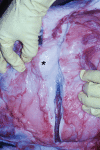
An 11-month-old, Holstein heifer was presented for a progressive swelling on the left side of the face that had been present since birth. A diagnosis of fibromatosis was made, based on macroscopic and microscopic examination of the abnormal infiltrative tissue. Because of the poor prognosis the animal was euthanized.
Fibromatose desmoide congénitale chez une taure Holstein. Une taure Holstein agée de 11 mois a été présentée pour une tuméfaction faciale gauche progressive et observée dès la naissance. Un diagnostic de fibromatose a été posé suite aux examens macroscopique et microscopique du tissu anormal infiltrant. L’animal a été euthanasié étant donné le sombre pronostic.
(Traduit par les auteurs)
Figures


Similar articles
-
Clinical and postmortem findings of pentalogy of Fallot in an 18-month-old Holstein heifer.J Vet Med Sci. 2019 Dec 5;81(11):1676-1679. doi: 10.1292/jvms.19-0147. Epub 2019 Oct 1. J Vet Med Sci. 2019. PMID: 31582644 Free PMC article.
-
[Picture report No. 5. Fibroma on the 3rd eyelid of a heifer].Dtsch Tierarztl Wochenschr. 1978 May 5;85(5):179. Dtsch Tierarztl Wochenschr. 1978. PMID: 348444 German. No abstract available.
-
Congenital aganglionosis in a 3-day-old Holstein calf.Can Vet J. 2005 Apr;46(4):342-4. Can Vet J. 2005. PMID: 15943121 Free PMC article.
-
Congenital infantile fibromatosis of the cheek: report of a rare case and differential diagnosis.Int J Oral Maxillofac Surg. 2011 Nov;40(11):1309-13. doi: 10.1016/j.ijom.2011.05.002. Epub 2011 Jun 11. Int J Oral Maxillofac Surg. 2011. PMID: 21658911 Review.
-
Bilateral breast fibromatosis: case report and review of the literature.J Surg Educ. 2011 Jul-Aug;68(4):320-5. doi: 10.1016/j.jsurg.2011.02.001. Epub 2011 Mar 25. J Surg Educ. 2011. PMID: 21708372 Review.
Cited by
-
Congenital neoplasms in cattle: a literature review and multi-institutional case series.J Vet Diagn Invest. 2025 Jul;37(4):559-573. doi: 10.1177/10406387251324512. Epub 2025 Mar 11. J Vet Diagn Invest. 2025. PMID: 40070027 Free PMC article. Review.
References
-
- Fletcher CDM. Soft tissue tumors. In: Fletcher CDM, editor. Diagnostic Histopathology of Tumors. 2. Vol. 2. London: Churchill Livingstone; 2000. pp. 1473–1540.
-
- Pool RR, Thompson KG. Tumors of joints. In: Meuten DJ, editor. Tumors in Domestic Animals. 4. Ames, Iowa: Blackwell Publ; 2002. pp. 240–241.
-
- Adams RD, editor. A Study in Pathology. 3. Hagerstown, Maryland: Harper and Row; 1975. Diseases of Muscle; pp. 389–390.
-
- Lever WF, Schaumburg-Lever G, editors. Histopathology of the Skin. 6. Philadelphia: JB Lippincott; 1983. p. 607.
-
- Wever ID, Cin PD, Fletcher CDM, et al. Cytogenetic, clinical, and morphological correlations in 78 cases of fibromatosis: Report from the CHAMP study group. Mod Pathol. 2000;13:1080–1085. - PubMed
Publication types
MeSH terms
LinkOut - more resources
Full Text Sources
