. 2008 Nov;4(11):e1000222.
doi: 10.1371/journal.ppat.1000222. Epub 2008 Nov 28.

Migratory dermal dendritic cells act as rapid sensors of protozoan parasites

Affiliations

Migratory dermal dendritic cells act as rapid sensors of protozoan parasites

Lai Guan Ng et al. PLoS Pathog. 2008 Nov.

Abstract

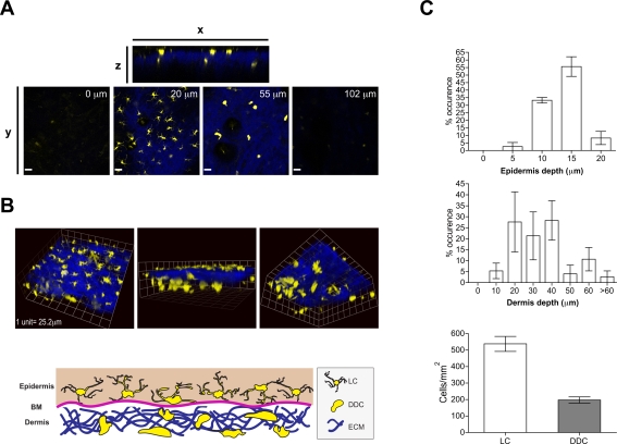
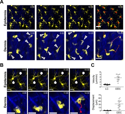
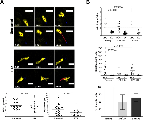
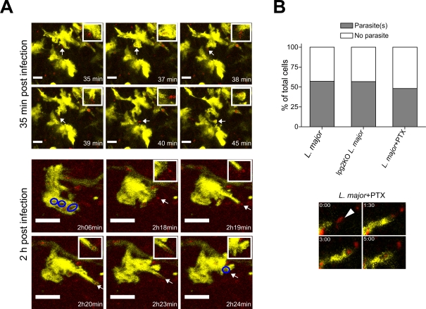
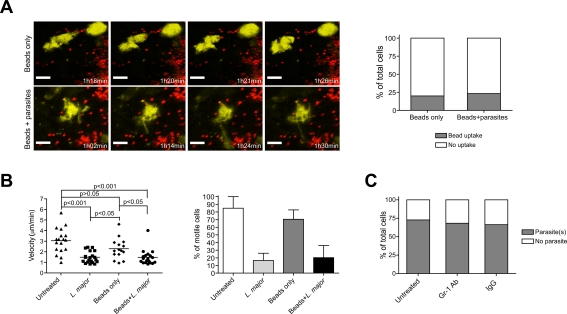
Dendritic cells (DC), including those of the skin, act as sentinels for intruding microorganisms. In the epidermis, DC (termed Langerhans cells, LC) are sessile and screen their microenvironment through occasional movements of their dendrites. The spatio-temporal orchestration of antigen encounter by dermal DC (DDC) is not known. Since these cells are thought to be instrumental in the initiation of immune responses during infection, we investigated their behavior directly within their natural microenvironment using intravital two-photon microscopy. Surprisingly, we found that, under homeostatic conditions, DDC were highly motile, continuously crawling through the interstitial space in a Galpha(i) protein-coupled receptor-dependent manner. However, within minutes after intradermal delivery of the protozoan parasite Leishmania major, DDC became immobile and incorporated multiple parasites into cytosolic vacuoles. Parasite uptake occurred through the extension of long, highly dynamic pseudopods capable of tracking and engulfing parasites. This was then followed by rapid dendrite retraction towards the cell body. DDC were proficient at discriminating between parasites and inert particles, and parasite uptake was independent of the presence of neutrophils. Together, our study has visualized the dynamics and microenvironmental context of parasite encounter by an innate immune cell subset during the initiation of the immune response. Our results uncover a unique migratory tissue surveillance program of DDC that ensures the rapid detection of pathogens.


Conflict of interest statement

The authors have declared that no competing interests exist.

Figures

Figure 1
Figure 1. Phenotypic characterization of CD11c-YFP+ cells in ear skin.
Flow cytometric analyses of surface markers expressed by epidermal and dermal cells from CD11c-YFP mice. The histogram plots were pre-gated on forward (FSC) and side-scatter (SSC) profiles. SSC/CD45 and FSC/YFP plots are shown for clear distinction of individual cell populations. Representative plots from 3 to 4 animals are shown.
Figure 2
Figure 2. Three-dimensional distribution of dendritic cells within CD11c-YFP mice.
(A) Single plane images from 2P-IVM showing YFP+ dendritic cells (yellow) in ear skin at various vertical depths along the z-projection. Extracellular matrix in the dermis was detected by the SHG signals (blue). Scale bar, 49 µm. (B) Upper panels, representative images from three-dimensional reconstructions of ear skin of a CD11c-YFP mouse showing the distribution of LC and DDC in relation to SHG. Lower panel, a schematic representation of DC localization in relation to different compartments in the skin. (C) Upper and middle histograms depict numbers of YFP+ LC and DDC along the vertical depth in the epidermis and dermis (underneath basement membrane). Lower histogram shows LC and DDC density in the epidermis and dermis (between 20–50 µm). Bars represent mean±SEM numbers obtained from at least three individual mice.
Figure 3
Figure 3. Migratory behavior of LC and DDC.
(A) Representative time-lapse images from 2P-IVM showing the migratory behavior of LC and DDC. Red line, track of migration during the observation period. Scale bar, 25 µm. (B) Representative high magnification time-lapse images showing the cellular movement of LC and DDC. Scale bars, 16 µm (epidermis) and 25 µm (dermis). Arrows illustrate dendrite movements. Red line, track of migration during the observation period. (C) Upper panel, mean velocity of LC and DDC; lower panel, displacement of LC and DDC from 15 min tracks. Symbols represent individual cells.
Figure 4
Figure 4. Migratory mechanisms of DDC and LC.
(A) Response of DDC to systemic injection of PTX. Top and middle panels depict a representative cell under control and PTX treatment conditions, respectively. The red square indicates the cell centroid, and the red line shows movement of the centroid over the observation period (n = 3 experiments for PTX treatment). Lower panel, data points represent individual cells, lines indicate mean. (B) Upper and middle plots show mean velocity and displacement of LC and DDC in response to systemic LPS challenge over time (n = 3 experiments). Data points represent individual cells, lines indicate mean. Lower plot shows the frequency of motile DDC at resting state, 2 to 4 h and 6 to 8 h after LPS treatment from 30 min tracks (bars represent mean±SEM).
Figure 5
Figure 5. Internalization of L. major by DDC.
(A) Three-dimensional reconstructions of ear skin inoculated with LmjF-DsRed2 promastigotes (red) showing the distribution of parasites (90 serial optical sections, 1 µm step size). (B) Representative images showing the morphology of LC (epidermis, yellow) and DDC (dermis, yellow) after LmjF-DsRed2 promastigote (red) inoculation. (C) A three-dimensional section of DDC (yellow) containing intracellular LmjF-DsRed2 promastigotes (red). Plot shows the frequency of LC and DDC containing LmjF or LV39 parasites (>50 cells obtained from randomly selected fields). (D) Comparison of the mean velocity and displacement of DDC in control skin, and DDC in infected skin with or without internalized parasites. Data points represent individual cells, lines indicate mean. Data were obtained from at least three independent experiments. (E) SNARF-1 was injected i.d. and DDC migration determined after 2 h (n = 3 experiments). Symbols represent individual cells. Control data are the same as in Figure 4A.
Figure 6
Figure 6. DDC extend pseudopods to engulf L. major parasites.
(A) Representative time-lapse images showing uptake of parasites (red) by rapid extension/retraction of pseudopods from DDC. Scale bars, 12 µm (upper panels) and 6 µm (lower panels). Small inlet shows tip of pseudopod at high magnification. Blue circles illustrate parasite-containing vacuoles. (B) Graph shows the effects of systemic PTX treatment on LmjLV39-DsRed2 parasite uptake by DDC in close vicinity to a parasite depot (n = 3 experiments). Images depict high magnification of parasite uptake by a dendrite after PTX treatment. Also shown is the uptake of lpg2KO-DsRed parasites by DDC (n = 3 experiments).
Figure 7
Figure 7. Discrimination of inert beads and L. major uptake by DDC and role of neutrophils in parasite uptake.
(A) Left panels, time-lapse images from 2P-IVM showing the cellular behaviors of DDC (yellow) after fluorescent bead inoculation alone (red) or beads (red) together with L. major promastigotes (unlabelled). Right panel, frequency of DDC with intracellular beads in the presence or absence of L. major parasites (>50 cells obtained from randomly selected fields). Scale bar, 12 µm. n = 3 experiments. (B) Mean velocity and frequency of motile DDC (mean±SEM ) at resting state, after LmjF-DsRed2 promastigote, beads only, and beads plus L. major promastigote inoculation. Data points represent individual cells. (C) Frequency of DDC with internalized LmjF-DsRed2 parasites in the ear skin of control (IgG), or neutrophil depleted (Gr-1 Ab) CD11c-YFP mice. n = 3 experiments.

Similar articles

Cited by

References

    1. Banchereau J, Steinman RM. Dendritic cells and the control of immunity. Nature. 1998;392:245–252. - PubMed
    1. Randolph GJ. Dendritic cell migration to lymph nodes: cytokines, chemokines, and lipid mediators. Semin Immunol. 2001;13:267–274. - PubMed
    1. Itano AA, McSorley SJ, Reinhardt RL, Ehst BD, Ingulli E, et al. Distinct dendritic cell populations sequentially present antigen to CD4 T cells and stimulate different aspects of cell-mediated immunity. Immunity. 2003;19:47–57. - PubMed
    1. Kissenpfennig A, Henri S, Dubois B, Laplace-Builhe C, Perrin P, et al. Dynamics and function of Langerhans cells in vivo: dermal dendritic cells colonize lymph node areas distinct from slower migrating Langerhans cells. Immunity. 2005;22:643–654. - PubMed
    1. Misslitz AC, Bonhagen K, Harbecke D, Lippuner C, Kamradt T, et al. Two waves of antigen-containing dendritic cells in vivo in experimental Leishmania major infection. Eur J Immunol. 2004;34:715–725. - PubMed

Publication types

Substances