. 2008;3(11):e3811.
doi: 10.1371/journal.pone.0003811. Epub 2008 Nov 26.

Lipopolysaccharide diversity evolving in Helicobacter pylori communities through genetic modifications in fucosyltransferases

Affiliations

Lipopolysaccharide diversity evolving in Helicobacter pylori communities through genetic modifications in fucosyltransferases

Christina Nilsson et al. PLoS One. 2008.

Abstract

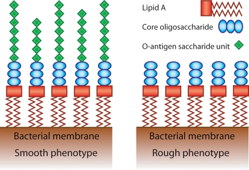
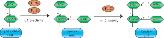
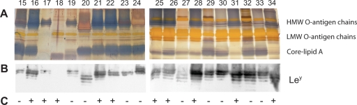
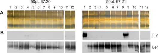
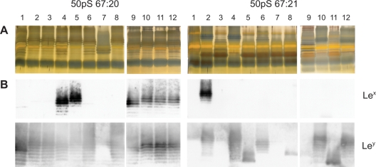
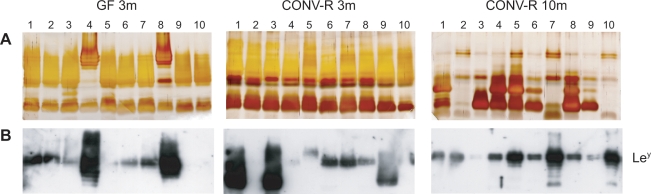
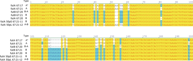
Helicobacter pylori persistently colonizes the gastric mucosa of half the human population. It is one of the most genetically diverse bacterial organisms and subvariants are continuously emerging within an H. pylori population. In this study we characterized a number of single-colony isolates from H. pylori communities in various environmental settings, namely persistent human gastric infection, in vitro bacterial subcultures on agar medium, and experimental in vivo infection in mice. The lipopolysaccharide (LPS) O-antigen chain revealed considerable phenotypic diversity between individual cells in the studied bacterial communities, as demonstrated by size variable O-antigen chains and different levels of Lewis glycosylation. Absence of high-molecular-weight O-antigen chains was notable in a number of experimentally passaged isolates in vitro and in vivo. This phenotype was not evident in bacteria obtained from a human gastric biopsy, where all cells expressed high-molecular-weight O-antigen chains, which thus may be the preferred phenotype for H. pylori colonizing human gastric mucosa. Genotypic variability was monitored in the two genes encoding alpha1,3-fucosyltransferases, futA and futB, that are involved in Lewis antigen expression. Genetic modifications that could be attributable to recombination events within and between the two genes were commonly detected and created a diversity, which together with phase variation, contributed to divergent LPS expression. Our data suggest that the surrounding environment imposes a selective pressure on H. pylori to express certain LPS phenotypes. Thus, the milieu in a host will select for bacterial variants with particular characteristics that facilitate adaptation and survival in the gastric mucosa of that individual, and will shape the bacterial community structure.


Conflict of interest statement

Competing Interests: The authors have declared that no competing interests exist.

Figures

Figure 1
Figure 1. Schematic representation of the LPS molecule.
The lipid A moiety consist of two parts, a lipid part composed by fatty acid chains that anchors LPS in the outer membrane and a sugar backbone that links to the core oligosaccharide. The outermost region of the LPS is built of repetitive saccharide units that form the O-antigen chain. The number of such units on a LPS molecule may vary within and between bacterial cells. In H. pylori these O-antigen saccharide units are usually structurally identical to Lewis antigens. For a detailed structure of H. pylori LPS, refer to references and . Bacteria that display the smooth LPS phenotype express all three moieties of the LPS molecule (left panel) whereas rough bacteria have lost the expression of the O-antigen chains (right panel).
Figure 2
Figure 2. Structures and synthetic pathway for Lewis antigens in H. pylori.
FutA and FutB can fucosylate Type 2 chains that are present in the elongating LPS molecule via an α1,3-FucT activity, creating the Lex antigen. Repetitive copies of such Lex units are usually present in the O-antigen chain of H. pylori. FutC that possesses an α1,2-FucT activity may subsequently add a second fucose to the Lex structure, generating the Ley antigen. This process will cap the O-antigen chain and block further elongation of the LPS molecule and, consequently, Ley can only be found at the terminal position of the O-antigen chain. Type 1 chains are structurally similar to the Type 2 chains and only differ in that they possess a β1,3-linkage instead of a β1,4-linkage between the Gal and GlcNAc saccharides in the precursor unit. In this case, fucosylation by FutA and FutB will require an α1,4-FucT activity and create a Lea antigen, prior to α1,2-fucosylation by FutC that produces a di-fucosylated Leb antigen. Gal = galactose, GlcNAc = N-Acetylglucosamine, Fuc = fucose.
Figure 3
Figure 3. LPS profiles of 20 intra-patient single-colony H. pylori isolates.
(A) Silver staining of extracted LPS from subisolates 67:15–34, illustrating the diversity of O-antigen chains within an H. pylori community in a persistent infection. The migration of high-molecular-weight (HMW) O-antigen chains, low-molecular-weight (LMW) O-antigen chains and core-lipid A is indicated. (B) Immunoblot analysis with antibodies detecting Ley reveals that all but one isolate express this antigen, but that the amount and size of Ley-bearing O-antigen chains differ among isolates. The area included in the immunoblot of part B corresponds to the HMW and LMW O-antigen regions in the silver stained gel of part A. (C) cag PAI status, as analyzed by PCR, is shown as + (present) or − (absent).
Figure 4
Figure 4. LPS profiles of H. pylori isolates after large bottleneck in vitro passages (50pL).
Silver staining (A) and Immunoblot analysis, detecting Lex and Ley (B), of extracted LPS from twelve single-colony isolates per strain, obtained after 50 in vitro passages of bacterial sweeps on agar medium, reveals minor differences in the LPS molecules.
Figure 5
Figure 5. LPS profiles of H. pylori isolates after small bottleneck in vitro passages (50pS).
Twelve colonies from 67:20 and 67:21 were initially isolated and passaged in parallel for 50 times on agar medium with one single colony being transferred in each subculturing step. Silver staining (A) and Immunoblot analysis, detecting Lex and Ley (B), of extracted LPS shows large variability in the O-antigen expression after such propagation, with loss of prominent high-molecular weight O-antigens in several of the 67:21-derived isolates..
Figure 6
Figure 6. LPS profiles of H. pylori isolates after in vivo passage in mice.
Strain 67:21 was used to infect one germ-free mouse for 3 months (GF 3m) and two conventionally raised mice for three (CONV-R 3m) and ten (CONV-R 10m) months, respectively. LPS was analyzed by silver staining (A) and immunoblot with antibodies detecting Ley (B) from ten emerging isolates from each mouse. The analyses revealed that the majority of isolates expressed LPS molecules with reduced levels of Ley-decorated high-molecular-weight O-antigens, as compared to the wildtype 67:21 strain.
Figure 7
Figure 7. Genetic exchange between futA and futB in H. pylori communities.
The 5′-region of futA and futB was sequenced in all isolates included in this study. The figure illustrates representative isolates that covers all nucleotide differences that were observed, the only exception being insertion/deletions of C-residues in the homopolymeric tract that varied according to table 1– 4.

References

    1. Mitchell HM, Li YY, Hu PJ, Liu Q, Chen M, et al. Epidemiology of Helicobacter pylori in southern China: identification of early childhood as the critical period for acquisition. J Infect Dis. 1992;166:149–153. - PubMed
    1. Granström M, Tindberg Y, Blennow M. Seroepidemiology of Helicobacter pylori infection in a cohort of children monitored from 6 months to 11 years of age. J Clin Microbiol. 1997;35:468–470. - PMC - PubMed
    1. Ernst PB, Gold BD. The disease spectrum of Helicobacter pylori: the immunopathogenesis of gastroduodenal ulcer and gastric cancer. Annu Rev Microbiol. 2000;54:615–640. - PubMed
    1. Suerbaum S, Michetti P. Helicobacter pylori infection. N Engl J Med. 2002;347:1175–1186. - PubMed
    1. Bourzac KM, Guillemin K. Helicobacter pylori-host cell interactions mediated by type IV secretion. Cell Microbiol. 2005;7:911–919. - PubMed

Publication types