. 2009 Feb;11(2):309-22.
doi: 10.1111/j.1462-5822.2008.01254.x. Epub 2008 Nov 28.

The mechanisms used by enteropathogenic Escherichia coli to control filopodia dynamics

Affiliations

The mechanisms used by enteropathogenic Escherichia coli to control filopodia dynamics

Cedric N Berger et al. Cell Microbiol. 2009 Feb.

Abstract

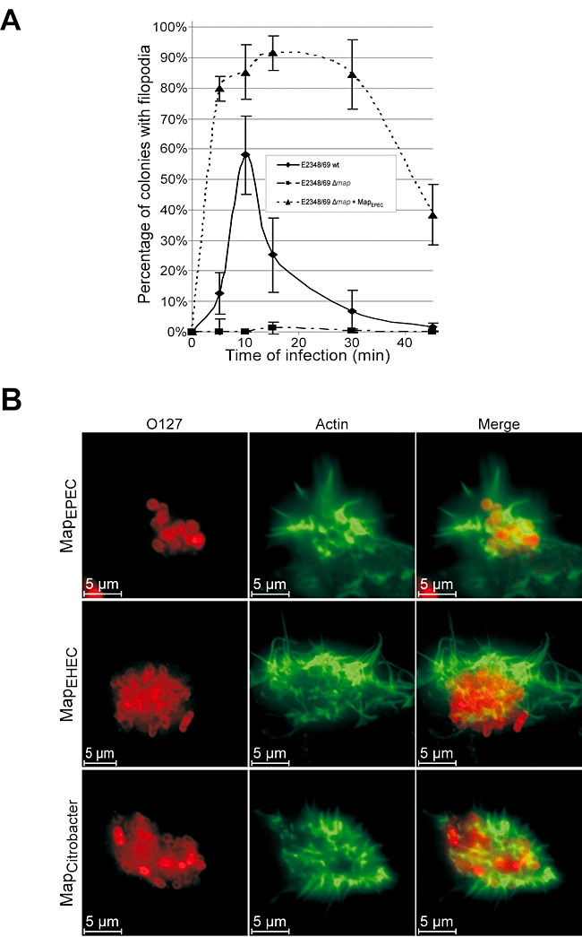
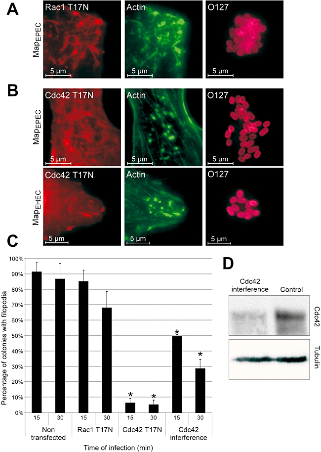
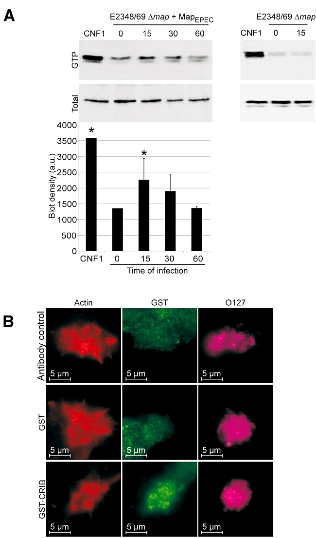
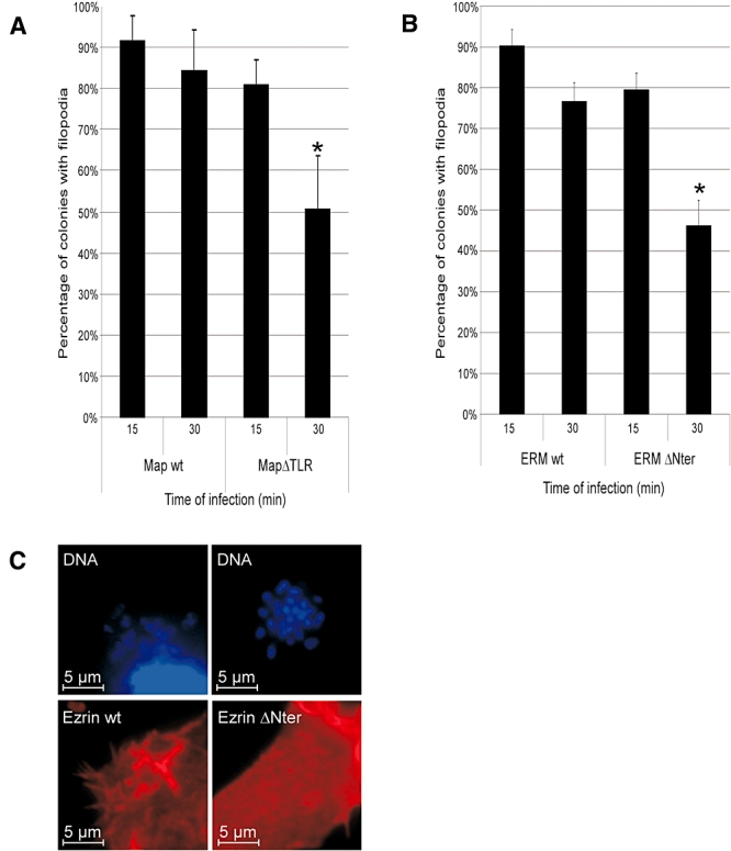
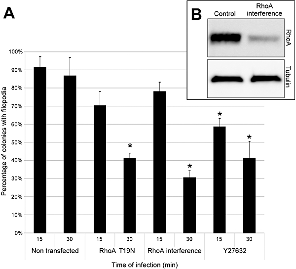
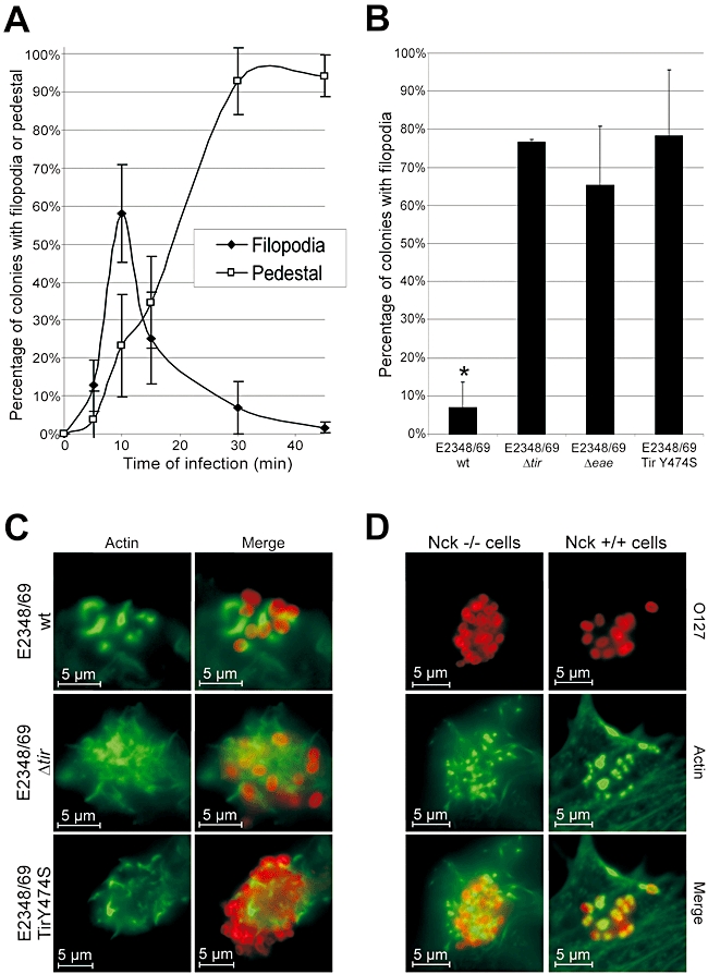
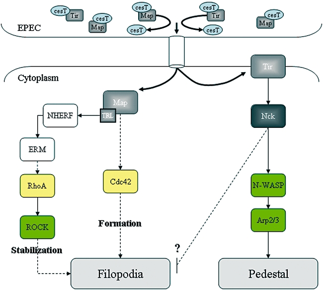
Enteropathogenic Escherichia coli (EPEC) subverts actin dynamics in eukaryotic cells by injecting effector proteins via a type III secretion system. First, WxxxE effector Map triggers transient formation of filopodia. Then, following recovery from the filopodial signals, EPEC triggers robust actin polymerization via a signalling complex comprising Tir and the adaptor proteins Nck. In this paper we show that Map triggers filopodia formation by activating Cdc42; expression of dominant-negative Cdc42 or knock-down of Cdc42 by siRNA impaired filopodia formation. In addition, Map binds PDZ1 of NHERF1. We show that Map-NHERF1 interaction is needed for filopodia stabilization in a process involving ezrin and the RhoA/ROCK cascade; expression of dominant-negative ezrin and RhoA or siRNA knock-down of RhoA lead to rapid elimination of filopodia. Moreover, we show that formation of the Tir-Nck signalling complex leads to filopodia withdrawal. Recovery from the filopodial signals requires phosphorylation of a Tir tyrosine (Y474) residue and actin polymerization pathway as both infection of cells with EPEC expressing TirY474S or infection of Nck knockout cells with wild-type EPEC resulted in persistence of filopodia. These results show that EPEC effectors modulate actin dynamics by temporal subverting the Rho GTPases and other actin polymerization pathways for the benefit of the adherent pathogen.


Figures

Fig. 1
Fig. 1
Kinetic of filopodia formation on 3T3 Swiss cells. A. Quantification of microcolony-associated filopodia on cell infected with wild-type E2348/69, E2348/69Δmap and E2348/69Δmap overexpressing MapEPEC. One hundred cells were counted in five independent experiments. Results are presented as mean ± SD. B. Fluorescence microscopy of 3T3 cells infected for 15 min with E2348/69Δmap overexpressing Map from EPEC, EHEC or C. rodentium. Actin was stained with Oregon green phalloidin (Green) and EPEC were detected with rabbit anti-0127 antibody (Red).
Fig. 2
Fig. 2
Filpodia formation by Map is Cdc42-dependent. 3T3 cells were transfected with dominant-negative Rac-1T17N (A) or Cdc42T17N (B) 24 h prior to infection with E2348/69Δmap overexpressing MapEPEC or MapEHEC for 15 min. Actin was stained with Oregon green phalloidin (Green), the Myc-tagged GTPases with mouse anti-myc (Red) and EPEC with rabbit anti-O127 (Magenta). Filopodia are observed on cells transfected with dominant-negative Rac-1 (A), but not on cells transfected with dominant-negative Cdc42 (B). Quantification of microcolony associated with filopodia in 3T3 cells transfected with dominant-negative Cdc42 or Rac-1 and Cdc42 siRNA (C). Cells were infected for 15 or 30 min with E2348/69Δmap overexpressing MapEPEC. One hundred colonies on transfected cell were counted in five independent experiments. Results are presented as mean ± SD. Significant differences from non-transfected cells are indicated by asterisks (*P < 0.01). Presence of filopodia induced by Map is affected by expression of Cdc42T17N or Cdc42 siRNA, but not by expression of Rac-1T17N. The level of Cdc42 and Tubulin in cell lysates 48 h after transfection with Cdc42 siRNA was determined by Western blots (D).
Fig. 3
Fig. 3
Map activates Cdc42. A. Swiss 3T3 cells were infected with E2348/69Δmap overexpressing MapEPEC for 15, 30 or 60 min. CNF1 toxin was used as a positive control (25 μg ml−1). Cells were lysed and a GST–CRIB fusion protein was used to co-purify Cdc42-GTP. Total Cdc42 in the lysates and Cdc42-GTP were detected by Western blotting with anti-Cdc42 antibodies. The graph shows measurement of blot densities from three pull-down experiments (means ± SD). Significant differences from uninfected cells (time 0) are indicated by asterisks (*P < 0.05). Infection with E2348/69Δmap did not induce Cdc42 activation. B. Fluorescence microscopy of 3T3 cells infected for 15 min with E2348/69Δmap overexpressing MapEPEC. Fixed cells were pre-incubated with purified GST or GST–CRIB. Actin was stained with TRITC phalloidin (Red), GST and GST–CRIB were detected with rabbit anti-GST antibody (Green) and EPEC were detected with goat anti-E. coli antibody (Magenta). Specific signals, at the base of filopodia, were observed in cell treated with GST–CRIB whereas no signal could be detected for control antibody or GST alone.
Fig. 4
Fig. 4
Filopodia stabilization is dependent on PDZ ligand motif (TRL) and ERM. Quantification of microcolony associated with filopodia on 3T3 cells (A) or 3T3 cell transfected with wild-type (wt) ezrin or dominant-negative ezrin (ΔNter) (B) infected for 15 or 30 min with E2348/69Δmap overexpressing MapEPEC (A and B) or MapΔTRL (A). One hundred colonies were counted from five independent experiments. Results are presented as mean ± SD. Significant differences are indicated by asterisks (*P < 0.01). Expression of MapΔTRL had no effect at 15 min, but resulted in reduced filopodia at 30 min post infection (A). Map-induced filopodia is affected by expression of ezrin ΔNter at 30 min post infection (B). Wild-type ezrin, but not ezrin ΔNter, is recruited to the Map-induced filopodia (C); VSV-G-tagged ERM was stained with rabbit anti-VSV-G (Red) and DNA with Hoechst (Blue).
Fig. 5
Fig. 5
Filopodia stabilization is dependent on RhoA. A. Quantification of microcolony associated with filopodia on 3T3 cells transfected with dominant-negative RhoAT19N and RhoA siRNA or pre-treated with ROCK inhibitor Y27632. Cells were infected for 15 or 30 min with E2348/69Δmap overexpressing MapEPEC. One hundred colonies on transfected cells were counted in five independent experiments. Results are presented as mean ± SD. Significant differences from non-transfected cells are indicated by asterisks (*P < 0.01). B. The level of RhoA and Tubulin in cell lysates 48 h after transfection with RhoA siRNA was determined on Western blots.
Fig. 6
Fig. 6
Downregulation of filopodia is dependent on Tir tyrosine phosphorylation and the Nck pathway. A. Quantification of microcolonies associated with filopodia or pedestals in 3T3 cell infected with wild-type E2348/69. One hundred cells were counted in five independent experiments. Results are presented as mean ± SD. B. Quantification of microcolonies associated with filopodia in cells infected for 30 min with E2348/69, E2348/69 Δtir, E2348/69 Δeae and E2348/69 expressing TirY474S. One hundred cells were counted in five independent experiments. Results are presented as mean ± SD. Significant differences from E2348/69 Δtir are indicated by asterisks (*P < 0.01). No filopodia were observed on cells infected with wild-type E2348/69 while filopodia were present on cell infected with E2348/69 Δtir, E2348/69 Δeae and E2348/69 expressing TirY474S. C. Fluorescent microscopy of 3T3 cells infected for 30 min with E2348/69, E2348/69 Δtir and E2348/69 TirY474S. Actin was stained with Oregon green phalloidin (Green) and EPEC were detected with rabbit anti-0127 antibody (Red). Pedestals are seen on cells infected with E2348/69 whereas filopodia are observed on cells infected with E2348/69 Δtir and E2348/69 expressing TirY474S. D. Wild-type or Nck-deficient MEF cells were infected for 15 min with E2348/69 Δmap overexpressing MapEPEC. Actin was stained with TRITC phalloidin (Red) and EPEC were detected with rabbit anti-0127 antibody (Magenta). Filopodia are seen on infected Nck-deficient MEF cells whereas pedestals are observed on infected control Nck+/+ cells.
Fig. 7
Fig. 7
A model of the mechanism used by EPEC to trigger filopodia formation and withdrawal. Prior to translocation Map and Tir are maintained in a translocation competent confirmation by association with the CesT chaperone (Creasey et al., 2003). Once translocated Map sequentially activates Cdc42 and RhoA. Activation of the Tir-Nck actin polymerization pathway leads to recovery from the filopodial signals (see text for further details).

References

    1. Algrain M, Turunen O, Vaheri A, Louvard D, Arpin M. Ezrin contains cytoskeleton and membrane binding domains accounting for its proposed role as a membrane-cytoskeletal linker. J Cell Biol. 1993;120:129–139. - PMC - PubMed
    1. Alto NM, Shao F, Lazar CS, Brost RL, Chua G, Mattoo S, et al. Identification of a bacterial type III effector family with G protein mimicry functions. Cell. 2006;124:133–145. - PubMed
    1. Arbeloa A, Bulgin RR, MacKenzie G, Shaw RK, Pallen MJ, Crepin VF, et al. Subversion of actin dynamics by EspM effectors of attaching and effacing bacterial pathogens. Cell Microbiol. 2008;10:1429–1441. - PMC - PubMed
    1. Arber S, Barbayannis FA, Hanser H, Schneider C, Stanyon CA, Bernard O, Caroni P. Regulation of actin dynamics through phosphorylation of cofilin by LIM-kinase. Nature. 1998;393:805–809. - PubMed
    1. Bladt F, Aippersbach E, Gelkop S, Strasser GA, Nash P, Tafuri A, et al. The murine Nck SH2/SH3 adaptors are important for the development of mesoderm-derived embryonic structures and for regulating the cellular actin network. Mol Cell Biol. 2003;23:4586–4597. - PMC - PubMed

Publication types

MeSH terms

LinkOut - more resources