. 2008 Dec;28(11):2213-20.
doi: 10.1111/j.1460-9568.2008.06514.x.

Calcitonin gene-related peptide (CGRP) triggers Ca2+ responses in cultured astrocytes and in Bergmann glial cells from cerebellar slices

Affiliations

Calcitonin gene-related peptide (CGRP) triggers Ca2+ responses in cultured astrocytes and in Bergmann glial cells from cerebellar slices

Stefano Morara et al. Eur J Neurosci. 2008 Dec.

Erratum in

  • Eur J Neurosci. 2008 Dec;28(12):2550

Abstract

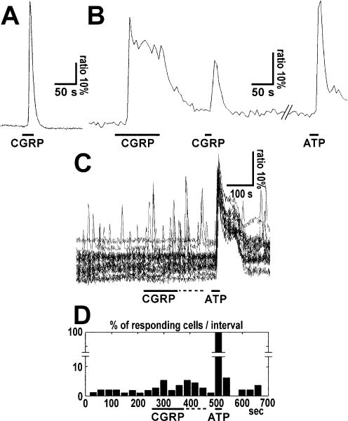
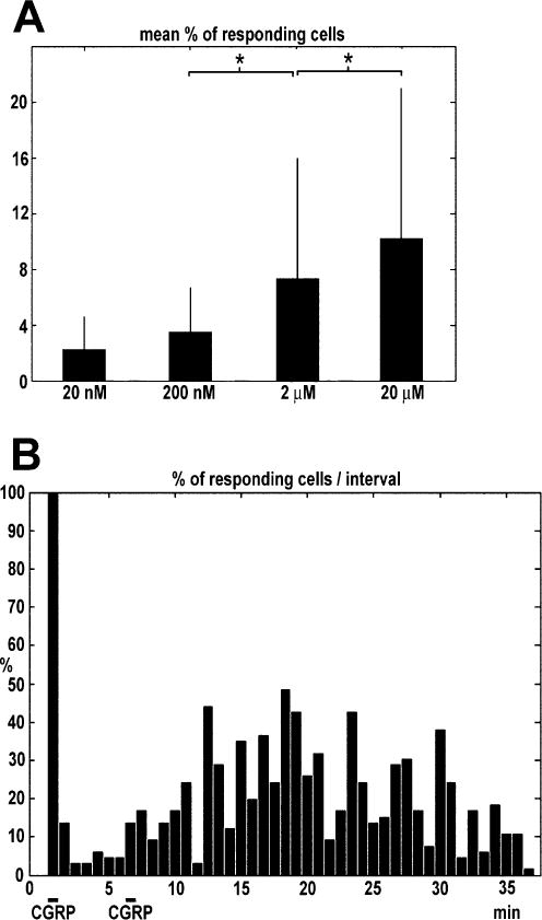
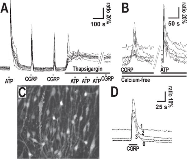
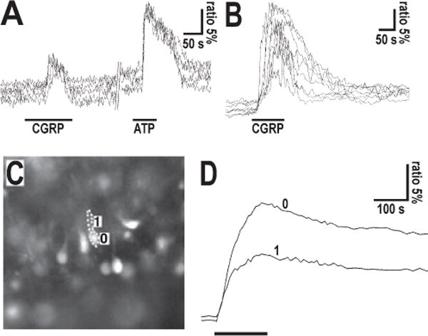
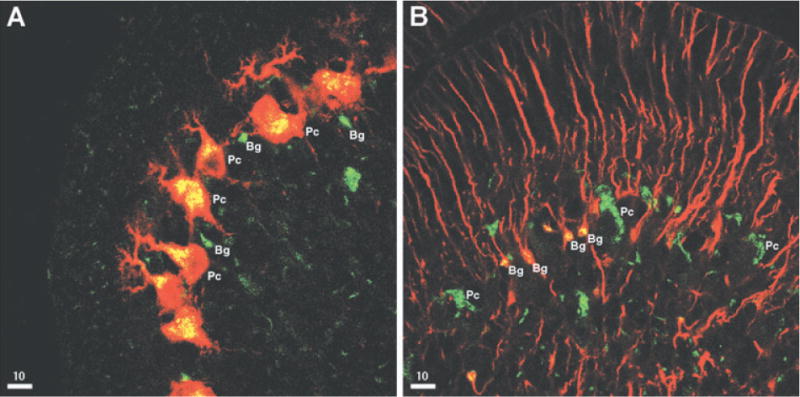
The neuropeptide calcitonin gene-related peptide (CGRP) is transiently expressed in cerebellar climbing fibers during development while its receptor is mainly expressed in astrocytes, in particular Bergmann glial cells. Here, we analyzed the effects of CGRP on astrocytic calcium signaling. Mouse cultured astrocytes from cerebellar or cerebral cortex as well as Bergmann glial cells from acutely isolated cerebellar slices were loaded with the Ca(2+) sensor Fura-2. CGRP triggered transient increases in intracellular Ca(2+) in astrocytes in culture as well as in acute slices. Responses were observed in the concentration range of 1 nm to 1 mm, in both the cell body and its processes. The calcium transients were dependent on release from intracellular stores as they were blocked by thapsigargin but not by the absence of extracellular calcium. In addition, after CGRP application a further delayed transient increase in calcium activity could be observed. Finally, cerebellar astrocytes from neonatal mice expressed receptor component protein, a component of the CGRP receptor, as revealed by immunofluorescence and confocal microscopy. It is thus proposed that the CGRP-containing afferent fibers in the cerebellum (the climbing fibers) modulate calcium in astrocytes by releasing the neuropeptide during development and hence possibly influence the differentiation of Purkinje cells.


Figures

FIG. 1
FIG. 1
Types of calcium responses induced in cultured cerebellar astrocytes by (A) pipette application of CGRP at medium to high concentrations or (B and C) by bath perfusion at low concentrations. (A) Simple transient calcium response to CGRP application. Ratiometric Fura-2 fluorescence recordings were used to determine changes in intracellular Ca2+ concentration. Note that the onset of decay occurred in the presence of the peptide. The bar indicates local pipette application of 250 nM CGRP. (B) Peak plus sustained plateau calcium responses in a single cultured cerebellar astrocyte after CGRP application. Note that the sustained calcium plateau was maintained in the presence of CGRP and Ca2+ returned to the basal level when the peptide was washed out. The bar indicates local pipette application of CGRP (50 μM) or ATP (100 μM) as control. (C) Ratiometric traces and (D) histogram of the percentage of responding cells per time interval are shown. CGRP (1 nM) and ATP (100 μM) were applied as indicated by bars. Only cells that responded at least once to CGRP were included. In D the columns represent the percentage of astrocytes which exhibited transient calcium responses in each 30 s time interval. The statistical analysis conducted by comparing two groups of seven time intervals corresponding to (i) the total CGRP period (from 240 to 450 s, i.e. CGRP application period plus the following 90 s) and (ii) the pre-CGRP application period (from 30 to 240 s) by means of the Wilcoxon signed-rank test showed that the increase in spike frequency was significant (P = 0.0313).
FIG. 2
FIG. 2
(A) Dose–response analysis of the effect of CGRP on astrocytic calcium and (B) biphasic response to CGRP. (A) Histogram representing the mean + SD percentage of responding cells recorded in 10 intervals (25 s each) during application of CGRP (20, 200 nM, 2 or 20 μM) by pipette in the bulk of the chamber solution. The mean background (spontaneous) calcium activity during 10 intervals before CGRP application has been subtracted. A statistical analysis of the comparison between spike frequencies recorded at different concentrations were carried out using the Kruskal–Wallis test, a nonparametric test for medians that was used because the samples did not follow a normal distribution. The test showed that medians were significantly different between 20 nM and 2 μM (P = 0.0270) and between 2 and 20 μM (P = 0.0217), whereas between 2 and 20 nM the difference was not significant (P = 0.9536). *P = 0.05. The number of experiments was 16 at each concentration. Numbers of cells: 20 nM, 426; 200 nM, 468; 2 μM, 407; 20 μM, 444. (B) The columns represent the percentage of astrocytes which exhibited transient calcium responses in each 30 s time interval. Note the two-phases type biphasic response the second application of CGRP. Bar indicates local pipette applications of CGRP (1 mM).
FIG. 3
FIG. 3
(A and B) Cellular mechanism and (C and D) localization of calcium responses to CGRP in cultured astrocytes. (A) Calcium responses of a cultured cerebrocortical astrocytic population to the application of CGRP before and after thapsigargin and ATP application. Ratiometric Fura-2 fluorescence recordings were used to determine changes in intracellular Ca2+ concentration. CGRP (10 μM), ATP (100 μM) and thapsigargin (125 μM) were locally applied by pipette as indicated by bars. (B) Calcium responses of a cultured cerebrocortical astrocytic population to the application of CGRP in Ca2+-free medium. CGRP was applied by pipette in the bulk of the chamber solution (away from cells) at 2 μM concentration and ATP at 4 μM, as indicated by bars. (C and D) Calcium responses in the soma and in processes from a single astrocyte cultured from cerebellum. In C the fluorescence image of the Fura-2-loaded astrocyte culture is shown. Four regions were selected (0, cell body; 1 and 2, proximal part of processes; 3, distal part of process) as indicated and the corresponding ratiometric Fura-2 fluorescence recordings are shown in D. Bar indicates 1 mm CGRP local application by pipette.
FIG. 4
FIG. 4
Calcium responses obtained in Bergmann glial cells from acute cerebellar slices. (A) Simple peak-type calcium responses in Fura-2-loaded Bergmann glial cells to local pipette application of CGRP (2.5 μM) and ATP (100 μM). Note that the onset of decay of the CGRP response occurreds in the presence of the peptide. The slice was prepared from a P4 mouse. (B) Peak plus plateau-type calcium responses in Bergmann glial cells to local pipette application of 50 μM CGRP. The slice was prepared from a P6 mouse cerebellum. Note that recovery from the plateau starts with cessation of CGRP application. (C, D) Calcium responses recorded from cell body and process of a single Bergmann glia cell from a cerebellar slice of a P3 mice. In (C) the fluorescence photograph of a Fura-2 loaded Bergmann glia cells is shown. The cell body and radial process (Bergmann glia fiber) of the analyzed cell has been separately outlined: 0 = cell body; 1 = radial process. In (D) the ratiometric traces of calcium concentration in the corresponding cell domains following local pipette application of CGRP (50 μM; indicated by bar) is shown.
FIG. 5
FIG. 5
Confocal images of immunoreactivity for RCP (green) and Calbindin D (A, red) or GFAP (B, red) from frontal sections of P7 cerebellum. Bergmann glia cell bodies (Bg) and Purkinje cell bodies (Pc) are indicated. The yellow color corresponds to colocalization.

References

    1. Adams JC, Mroz EA, Sewell WF. A possible neurotransmitter role for CGRP in a hair-cell sensory organ. Brain Res. 1987;419:347–351. - PubMed
    1. Aiyar N, Disa J, Stadel JM, Lysko PG. Calcitonin gene-related peptide receptor independently stimulates 3′,5′-cyclic adenosine monophosphate and Ca2+ signaling pathways. Mol Cell Biochem. 1999;197:179–185. - PubMed
    1. Altman J. Morphological development of the rat cerebellum and some of its mechanisms. In: Palay SL, Chan-Palay V, editors. The Cerebellum New Vistas. Springer Verlag; Berlin-Heidelberg: 1982. pp. 8–46.
    1. Armengol JA, Sotelo C. Early dendritic development of Purkinje cells in the rat cerebellum. A light and electron microscopic study using axonal tracing in ‘in vitro’ slices. Dev Brain Res. 1991;64:95–114. - PubMed
    1. Burns DM, Stehno-Bittel L, Kawase T. Calcitonin gene-related peptide elevates calcium and polarizes membrane potential in MG-63 cells by both cAMP-independent and -dependent mechanisms. Am J Physiol Cell Physiol. 2004;287:C457–C467. - PubMed

Publication types

MeSH terms