. 2008 Dec 1:5:17.
doi: 10.1186/1743-8977-5-17.

Pulmonary response to intratracheal instillation of ultrafine versus fine titanium dioxide: role of particle surface area

Affiliations

Pulmonary response to intratracheal instillation of ultrafine versus fine titanium dioxide: role of particle surface area

Tina M Sager et al. Part Fibre Toxicol. .

Abstract

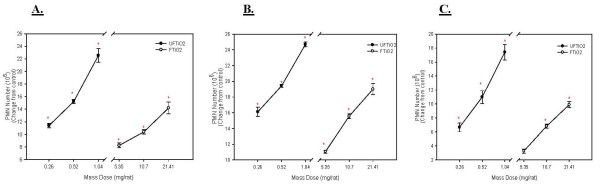
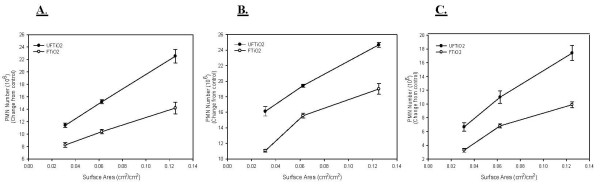
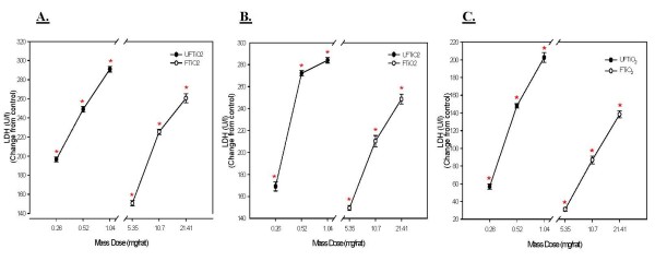
Background: The production and use of nanoparticles is growing rapidly due to the unique physical and chemical properties associated with their nano size and large surface area. Since nanoparticles have unique physicochemical properties, their bioactivity upon exposure to workers or consumers is of interest. In this study, the issue of what dose metric (mass dose versus surface area dose) is appropriate for toxicological studies has been addressed. Rats were exposed by intratracheal instillation to various doses of ultrafine or fine TiO2. At 1, 7, or 42 days post-exposure, inflammatory and cytotoxic potential of each particle type was compared on both a mass dosage (mg/rat) as well as an equal surface area dosage (cm2 of particles per cm2 of alveolar epithelium) basis.

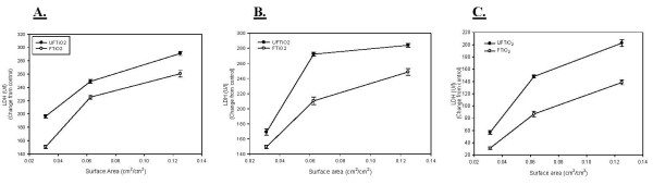
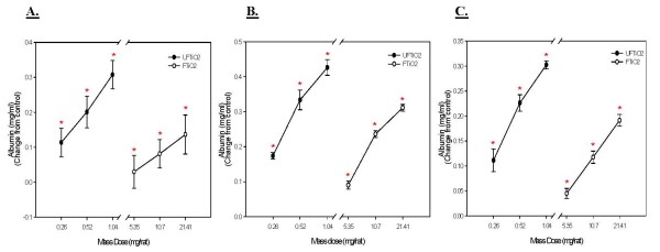
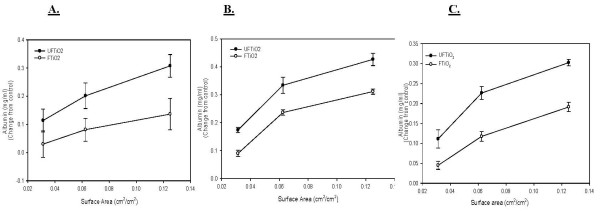
Results: The findings of the study show that on a mass basis the ultrafine particles caused significantly more inflammation and were significantly more cytotoxic than the fine sized particles. However, when doses were equalized based on surface area of particles delivered, the ultrafine particles were only slightly more inflammogenic and cytotoxic when compared to the fine sized particles. Lung burden data indicate that ultrafine TiO2 appears to migrate to the interstitium to a much greater extent than fine TiO2.

Conclusion: This study suggests that surface area of particles may be a more appropriate dose metric for pulmonary toxicity studies than mass of particles.


Figures

Figure 1
Figure 1
Electron micrographs of titanium dioxide particles suspended in BALF. A transmission electron micrograph of UFTiO2 suspended in BALF (A). Magnification of 60,000 × (note 500 nm scale bar). A scanning electron micrograph of FTiO2 suspended in BALF (B). Magnification of 20,000 × (note 1 um scale bar).
Figure 2
Figure 2
A comparison of inflammation elicited in animals receiving various mass doses of UFTiO2 and FTiO2 suspended in BALF. A comparison of inflammation elicited in animals receiving various mass doses of UFTiO2 and FTiO2 suspended in BALF at 1 day (Panel A), 7 days (Panel B), and 42 days (Panel C) post-exposure. Rats were exposed to various mass doses of UFTiO2 and FTiO2 by intratracheal instillation. Animals were euthanized at 1 day, 7 days, and 42 days post-exposure and bronchoalveolar lavage was performed. Inflammation was assessed by BAL PMN counts. Values are increased PMN number above the BALF control and are given as means ± SE of 8 experiments. Control PMN values were 1.37 ± 0.098 × 10⁶, 0.78 ± 0.074 × 10⁶, and 0.88 ± 0.095 × 10⁶ cells/rat for 1, 7 and 42 days, respectively. Linear regression analysis with a 95% confidence interval reveals that on a mass dose basis UFTiO2 causes significantly more inflammation than FTiO2 at all post-exposure time points. * indicates a significant increase from control (p < 0.05; ANOVA).
Figure 3
Figure 3
A comparison of inflammation elicited in animals receiving doses of UFTiO2 and FTiO2 normalized to surface area of particles administered per surface area of alveolar epithelium. A comparison of inflammation elicited in animals receiving doses (0.0313, 0.0625 and 0.125 cm2/cm2) of UFTiO2 and FTiO2 normalized to surface area of particles administered per surface area of alveolar epithelium at 1 day (Panel A), 7 days (Panel B), and 42 days (Panel C) post-exposure. Particles were suspended in BALF. Rats were exposed to various doses of UFTiO2 and FTiO2 by intratracheal instillation. Animals were euthanized at 1 day, 7 days, and 42 days post-exposure and bronchoalveolar lavage was performed. Inflammation was assessed by BAL PMN counts. Values are increased PMN number above the BALF control and are given as means ± SE of 8 experiments. Control PMN values were 1.37 ± 0.098 × 10⁶, 0.78 ± 0.074 × 10⁶, and 0.88 ± 0.095 × 10⁶ cells/rat for 1, 7 and 42 days, respectively. Linear regression analysis with a 95% confidence interval reveals that, when dose is normalized to surface area of particles administered, dose responses curves assessing inflammation caused by UFTiO2 and FTiO2 exposure are not significantly different. On a dose normalized to surface area, UFTiO2 elicits at most a 2 fold increase in inflammation when compared to FTiO2 at all post-exposure times.
Figure 4
Figure 4
A comparison of cellular cytotoxicity elicited in animals receiving various mass doses of UFTiO2 and FTiO2 suspended in BALF. A comparison of cellular cytotoxicity elicited in animals receiving various mass doses of UFTiO2 and FTiO2 suspended in BALF at 1 day (Panel A), 7 days (Panel B), and 42 days (Panel C) post-exposure. Rats were exposed to various mass doses of UFTiO2 and FTiO2 by intratracheal instillation. Animals were euthanized at 1 day, 7 days, and 42 days post-exposure and bronchoalveolar lavage was performed. Cytotoxicity was assessed by measuring LDH activity. Values are increase in LDH activity above the BALF control and are given as means ± SE of 8 experiments. Control values of LDH activity were 46.375 ± 2.24, 39.5 ± 1.35, and 37.25 ± 2.63 U/l for 1, 7 and 42 days, respectively. Linear regression analysis with a 95% confidence interval reveals that on a mass dose basis UFTiO2 causes significantly more LDH activity than FTiO2 at all post-exposure time points. * indicates a significant increase from control (p < 0.05; ANOVA).
Figure 5
Figure 5
A comparison of cytotoxicity elicited in animals receiving doses of UFTiO2 and FTiO2 normalized to surface area of particle administered per surface area of alveolar epithelium. A comparison of cytotoxicity elicited in animals receiving doses (0.0313, 0.0625 and 0.125 cm2/cm2) of UFTiO2 and FTiO2 normalized to surface area of particles administered per surface area of alveolar epithelium at 1 day (Panel A), 7 days (Panel B), and 42 days (Panel C) post-exposure. Animals were exposed to various doses of UFTiO2 and FTiO2 by intratracheal instillation. Animals were euthanized at 1 day, 7 days, and 42 days post-exposure and bronchoalveolar lavage was performed. Cytotoxicity was assessed by measuring LDH activity. Values are increase in LDH activity above the BALF control and are given as means ± SE of 8 experiments. Control values of LDH activity were 46.375 ± 2.24, 39.5 ± 1.35, and 37.25 ± 2.63 U/l for 1, 7 and 42 days, respectively. Linear regression analysis with 95% confidence internal reveals that, when dose is normalized to surface area of particles administered, responses to UFTiO2 and FTiO2 are not significantly different.
Figure 6
Figure 6
A comparison of cellular damage elicited in animals receiving various mass doses of UFTiO2 and FTiO2 suspended in BALF. A comparison of cellular damage elicited in animals receiving various mass doses of UFTiO2 and FTiO2 suspended in BALF at 1 day (Panel A), 7 days (Panel B), and 42 days (panel C) post-exposure. Rats were exposed to various mass doses of UFTiO2 and FTiO2 by intratracheal instillation. Animals were euthanized at 1 day, 7 days, and 42 days post-exposure and bronchoalveolar lavage was performed. Cellular injury was assessed by measuring albumin levels. Control values of albumin levels were 0.073 ± 0.033, 0.084 ± 0.003, 0.098 ± 0.007 mg/ml for 1, 7, and 42 days, respectively. Values are increase in albumin levels above the BALF control and are given as means ± SE of 8 experiments. * indicates a significant increase from control (p < 0.05; ANOVA).
Figure 7
Figure 7
A comparison of cellular damage elicited in animals receiving doses of UFTiO2 and FTiO2 normalized to surface area of particle administered per surface area of alveolar epithelium. A comparison of cellular damage elicited in animals receiving doses (0.0313, 0.0625 and 0.125 cm2/cm2) of UFTiO2 and FTiO2 normalized to surface area of particles administered per surface area of alveolar epithelium at 1 day (Panel A), 7 days (Panel B), and 42 days (Panel C) post-exposure. Rats were exposed to various mass doses of UFTiO2 and FTiO2 by intratracheal instillation. Animals were euthanized at 1 day, 7 days, and 42 days post-exposure and bronchoalveolar lavage was performed. Cellular injury was assessed by measuring albumin levels. Control values of albumin levels were 0.073 ± 0.033, 0.084 ± 0.003, 0.098 ± 0.007 mg/ml for 1, 7, and 42 days, respectively. Values are increase in albumin levels above the BALF control and are given as means ± SE of 8 experiments. Linear regression analysis with a 95% confidence interval reveals that, when dose is normalized to surface area of particles administered, responses to UFTiO2 and FTiO2 are not significantly differently.

References

    1. McAllister K, Sazani P, Adam M, Cho M, Rubinstein M, Samuluski R. Polymeric nanogels produced via inverse microemulsion polymerization as potential gene and antisense delivery agents. J Am Chem Soc. 2002;124:15198–15207. doi: 10.1021/ja027759q. - DOI - PubMed
    1. Grassian V, O'Shaughnessy P, Adamcakova-Dodd A, Pettibone J. Inhalation exposure study of titanium dioxide nanoparticles with a primary particle size of 2 to 5 nm. Environ Health Perspect. 2007;115:397–402. - PMC - PubMed
    1. Donaldson K, Li XY, MacNee W. Ultrafine (nanometer) particle mediated lung injury. J Aerosol Sci. 1998;29:553–60. doi: 10.1016/S0021-8502(97)00464-3. - DOI
    1. Oberdorster G. Significance in parameters in the evaluation of exposure-dose response relationships of inhaled particles. Inhal Toxicol. 2002. pp. 73–89. - PubMed
    1. Maynard A, Zimmer T. Evaulation of grinding aerosols in terms of alveolar dose: the significance of using mass, surface area and number metrics. Ann Occup Hyg. 2002;46:315–319.

LinkOut - more resources