. 2008 Dec 1:1:122.
doi: 10.1186/1756-0500-1-122.

Resveratrol prevents antibody-induced apoptotic death of retinal cells through upregulation of Sirt1 and Ku70

Affiliations

Resveratrol prevents antibody-induced apoptotic death of retinal cells through upregulation of Sirt1 and Ku70

Thimmappa S Anekonda et al. BMC Res Notes. .

Abstract

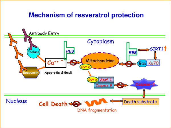
Background: To determine whether resveratrol, a natural plant-derived drug, has protective effects against antibody-induced apoptosis of retinal cells in vitro and to provide insights on the mechanism of resveratrol protection.

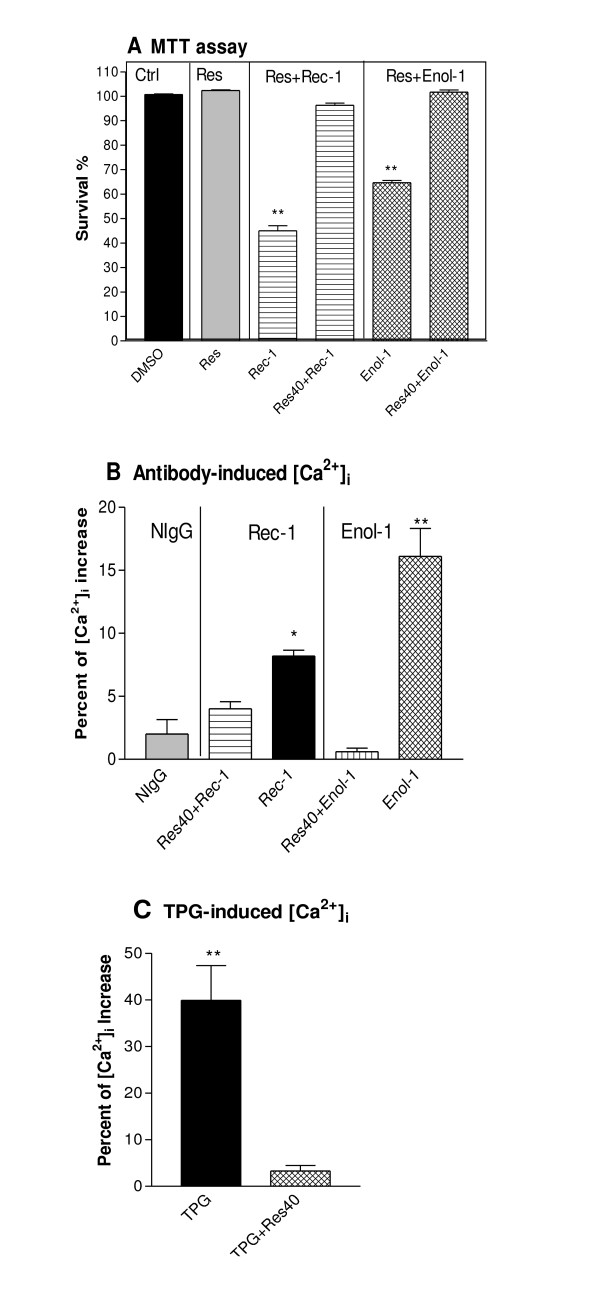
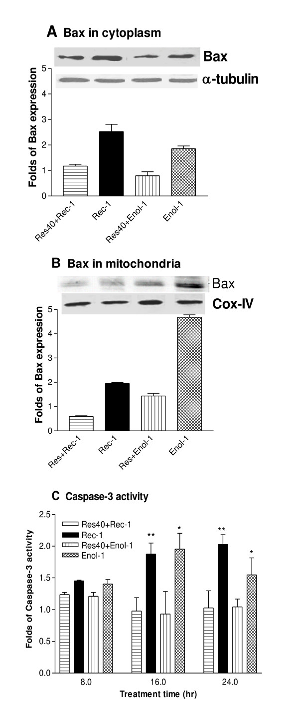
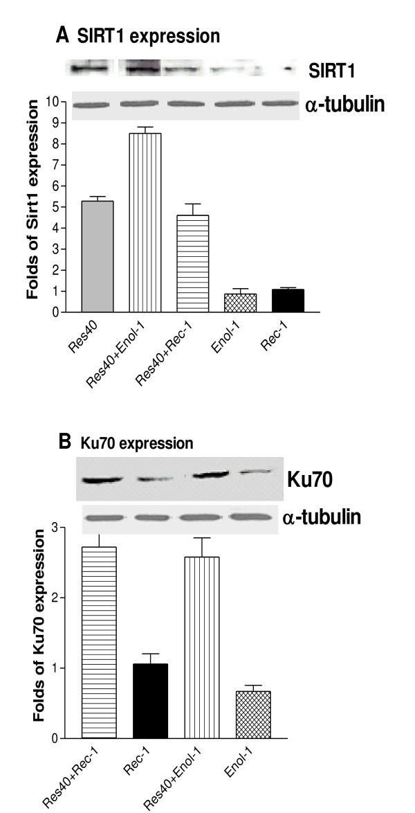
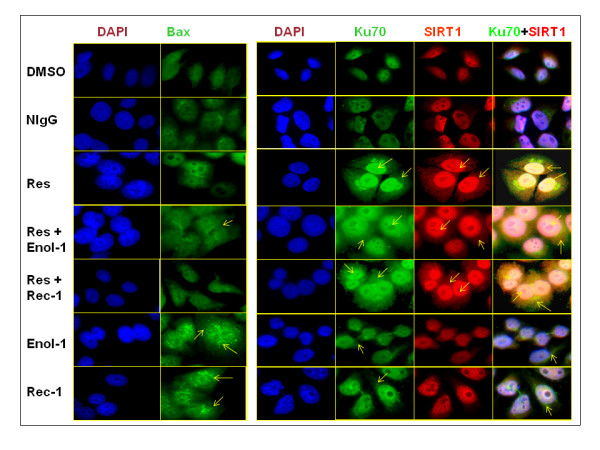
Findings: E1A.NR3 retinal cells pretreated with 40 muM resveratrol were grown in the presence of anti-recoverin (Rec-1), anti-enolase (Enol-1) antibodies, and normal purified immunoglobulins. When the cells were exposed to resveratrol before treatment with Enol-1 or Rec-1 antibodies, 30-55% more cells survived compared to the resveratrol-untreated cells. Western blotting showed a reduction in proapoptotic protein Bax in the cytoplasm and mitochondria of resveratrol-treated cells. Resveratrol-pretreated cells also showed a significant decrease in intracellular calcium and an inhibition of caspase-3 activity as compared to the untreated cells. Sirt1 expression was greatly reduced in the cells grown in the presence of Rec-1 and Enol-1, but it increased about five times in the resveratrol-pretreated cells. Immunocytochemistry revealed that Sirt1 expression in the cytoplasm and nucleus was colocalized with Ku70 expression in resveratrol-treated cells, suggesting possible interaction with each other in the cell. The pattern of the Ku70 cellular localization also overlapped with the Bax cellular localization in treated and untreated cells.

Conclusion: In vitro protection of retinal cells from apoptosis by resveratrol occurred through multiple early molecular events, such as reduction of intracellular calcium levels, down-regulation of Bax, up-regulation of Sirt1 and Ku70 activities, and inhibition of caspase-3 activity. These findings will help designing future in vivo and pre-clinical treatments for autoimmune retinopathies.


Figures

Figure 1
Figure 1
Protection of retinal cells by resveratrol against antibody-induced cytotoxicity. (A) E1A.NR3 cells were grown in 96-well plates (100 μl) for 72 hr in the presence of 0.8 mg/ml purified Rec-1 or Enol-1 alone, or antibodies plus 40 μM resveratrol (Res). Cell survival was measured by MTT assay. Each treatment was replicated three times, and the bars represent an average percentage of cell survival for 3 experiments. The statistical significance of each treatment was estimated against controls using one-way ANOVA and Mann-Whitney t-tests. ** = p < 0.01. (B) Effects of resveratrol on antibody-induced intracellular [Ca2+] levels. Cells were incubated in a fluorescent dye for 30 min before the addition of 50 μl of 0.8 mg/ml Rec-1 and Enol-1 or (C) 2 μM thapsigargin (TPG), individually or after 15 min pre-treatment with 40 μM resveratrol. Each treatment was replicated three times and the experiment was repeated thrice. The statistical significance of each treatment was estimated against DMSO controls using one-way ANOVA and Mann-Whitney t-tests. * = p ≤ 0.05; ** = p ≤ 0.01.
Figure 2
Figure 2
Molecular effects of resveratrol on pro-apoptotic proteins in E1A.NR3 retinal cells. Effects of resveratrol on Bax expression in the cytoplasm (A) and mitochondria (B). The retinal cells were treated with 40 μM resveratrol (Res) for 16 hrs followed by treatment with 0.8 mg/ml of Rec-1 and Enol-1 for 45 min. The protein expression was determined by Western blotting and the expression level is presented as a fold increase calculated from the intensity of each band compared to the untreated control after adjusting for the loading control using densitometry analyses. The bars represent an average of three experiments. (C) Effects of resveratrol on caspase-3 activity. The EnzChek Caspase-3 fluorescent assay was used to determine the protease activity of caspase-3 in E1A.NR3 cells in the presence and absence of 40 μM resveratrol pretreatment (16 hrs) followed by induction of apoptosis using 0.8 mg/ml of Rec-1 or Enol-1 for 8, 16, and 24 hrs. Each treatment was replicated three times and the experiment was repeated twice. The statistical significance of treatments under each time of exposure was estimated against vehicle control using two-way ANOVA and Mann-Whitney t-tests. * = p ≤ 0.05; ** = p ≤ 0.01.
Figure 3
Figure 3
Molecular effects of resveratrol on anti-apoptotic proteins. Effects of resveratrol on (A) Sirt1 and (B) Ku70 expression in antibody-treated E1A-NR3 cells. The cells were pretreated with 40 μM resveratrol for 16 hrs followed by 2 hrs incubation in Enol-1 or Rec-1 antibodies. The protein cell extracts were analyzed by Western blotting using anti-Sirt1 and anti-Ku70 antibodies. Expression level is presented as a fold increase calculated from the intensity of each band compared to the untreated controls adjusted for the loading control using a densitometry analyses. The bars represent an average of three experiments.
Figure 4
Figure 4
Immunofluorescent analysis of pro-and anti-apoptotic proteins. Immunofluorescent analysis of the expression of Bax (green), Ku70 (green), and Sirt1 (red) proteins in E1A.NR3 retinal cells in the presence and absence of 40 μM resveratrol pretreatment (16 hrs) followed by induction of apoptosis using 0.8 mg/ml of Rec-1 or Enol-1 for 2 hrs. The cells were incubated with anti-Bax, anti-Ku70 and anti-Sirt1 antibodies followed by incubation in appropriate fluorescent secondary antibodies. Nuclei were stained with DAPI (blue) for 10 min. The images were photographed under a Leica DM5000B fluorescence microscope. Each treatment was replicated three times and the experiment was repeated twice. The arrows indicate the expression of corresponding proteins in the cytoplasm of nucleus.
Figure 5
Figure 5
A proposed molecular mechanism of resveratrol protection against antibody-induced apoptosis in retinal cells. Antibodies enter the cell through endocytosis and induce an increase in intracellular calcium levels, which in turn trigger the mitochondria-mediated increase in caspase 3 activity and apoptotic death of retinal cells. Resveratrol blocked the intracellular calcium level and blocked the entry of Bax from cytoplasm to mitochondria, which subsequently short-circuited the caspase-3 activation and the cell from death. This figure has been modified from a previously published figure in [9].

References

    1. Keltner JL, Thirkill CE, Tyler NK, Roth AM. Management and monitoring of cancer-associated retinopathy. Arch Ophthalmol. 1992;110:48–53. - PubMed
    1. Congdon N, O'Colmain B, Klaver CC, Klein R, Munoz B, Friedman DS, Kempen J, Taylor HR, Mitchell P. Causes and prevalence of visual impairment among adults in the United States. Arch Ophthalmol. 2004;122:477–485. doi: 10.1001/archopht.122.4.477. - DOI - PubMed
    1. Weleber RG, Watzke RC, Shults WT, Trzupek KM, Heckenlively JR, Egan RA, Adamus G. Clinical and electrophysiologic characterization of paraneoplastic and autoimmune retinopathies associated with antienolase antibodies. Am J Ophthalmol. 2005;139:780–794. doi: 10.1016/j.ajo.2004.12.104. - DOI - PubMed
    1. Thirkill CE, Roth AM, Keltner JL. Cancer-associated retinopathy. Arch Ophthalmol. 1987;105:372–375. - PubMed
    1. Adamus G, Ren G, Weleber RG. Autoantibodies against retinal proteins in paraneoplastic and autoimmune retinopathy. BMC Ophthalmol. 2004;4:5. doi: 10.1186/1471-2415-4-5. - DOI - PMC - PubMed