. 2009 Mar 20;284(12):7561-77.
doi: 10.1074/jbc.M807497200. Epub 2008 Dec 1.

Role of Src signal transduction pathways in scatter factor-mediated cellular protection

Affiliations

Role of Src signal transduction pathways in scatter factor-mediated cellular protection

Saijun Fan et al. J Biol Chem. .

Abstract

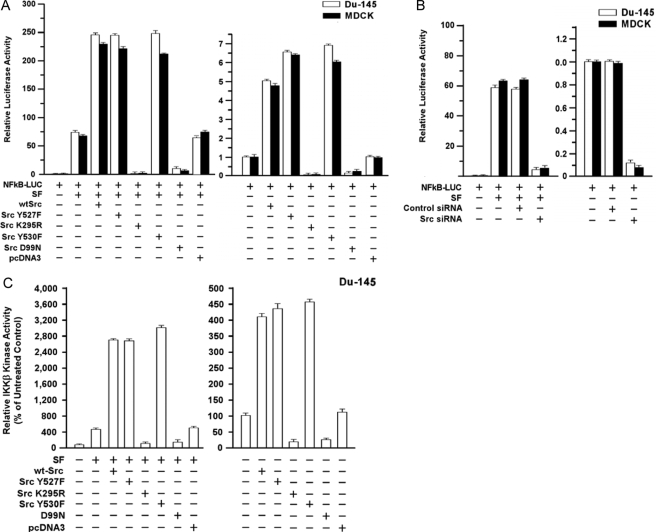
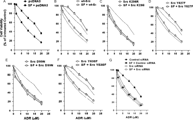
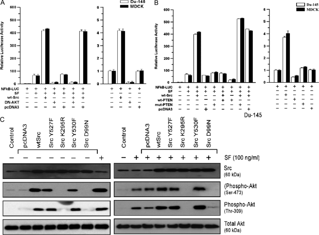
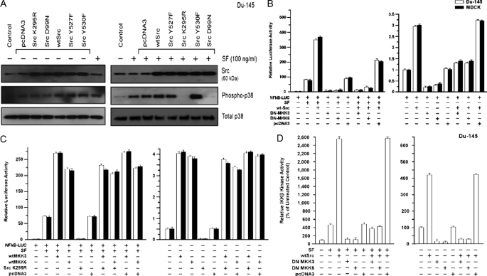
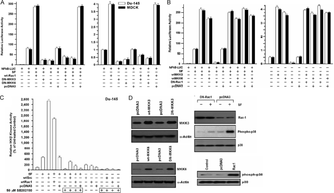
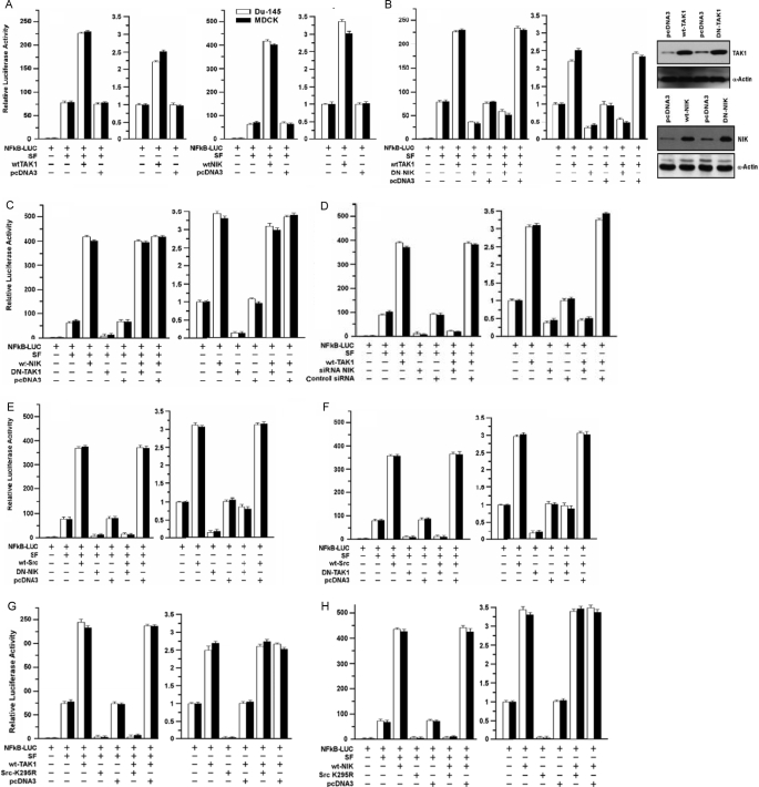
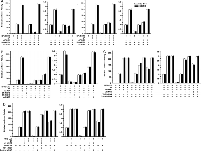
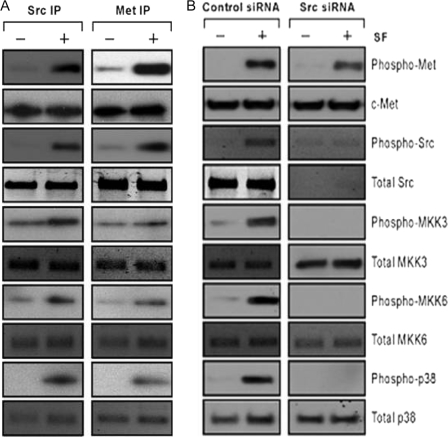
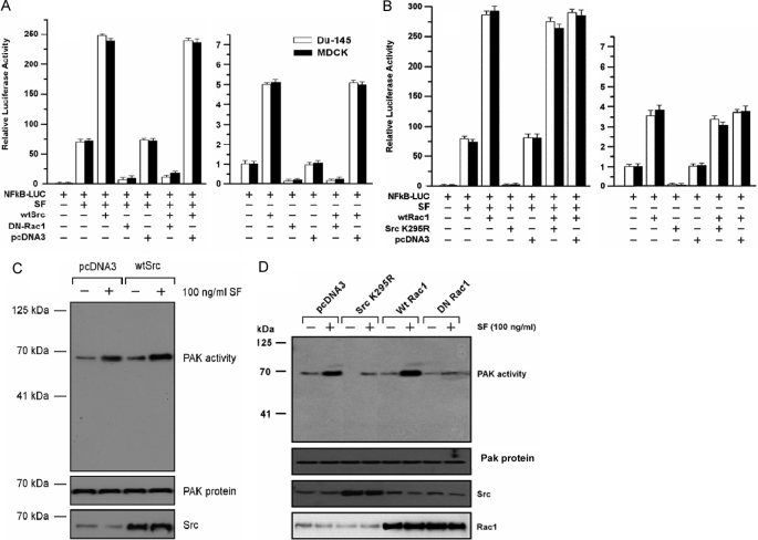
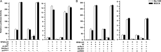
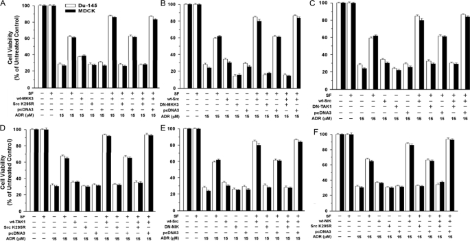
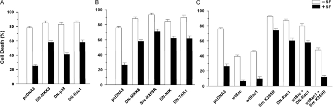
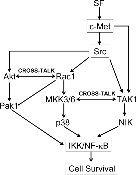
Scatter factor (SF) (hepatocyte growth factor) is a pleiotrophic cytokine that accumulates in tumors, where it may induce invasion, angiogenesis, and chemoresistance. We have studied the mechanisms by which SF and its receptor (c-Met) protect cells against the DNA-damaging agent adriamycin (ADR) as a model for chemoresistance of SF/c-Met-overexpressing tumors. Previous studies identified a phosphatidylinositol 3-kinase/c-Akt/Pak1/NF-kappaB cell survival pathway in DU-145 prostate cancer and Madin-Darby canine kidney epithelial cells. Here we studied Src signaling pathways involved in SF cell protection. Src enhanced basal and SF stimulated NF-kappaB activity and SF protection against ADR, in a manner dependent upon its kinase and Src homology 3 domains; and endogenous Src was required for SF stimulation of NF-kappaB activity and cell protection. The ability of Src to enhance SF stimulation of NF-kappaB activity was due, in part, to its ability to stimulate Akt and IkappaB kinase activity; and Src-mediated stimulation of NF-kappaB was due, in part, to a Rac1/MKK3/6/p38 pathway and was Akt-dependent. SF caused the activation of Src and the Rac1 effector Pak1. Furthermore, SF induced activating phosphorylations of MKK3, MKK6, and p38 within the c-Met signalsome in an Src-dependent manner. The NF-kappaB-inducing kinase was found to act downstream of TAK1 (transforming growth factor-beta-activated kinase 1) as a mediator of SF- and Src-stimulated NF-kappaB activity. Finally, the Src/Rac1/MKK3/6/p38 and Src/TAK1/NF-kappaB-inducing kinase pathways exhibited cross-talk at the level of MKK3. These findings delineate some novel signaling pathways for SF-mediated resistance to ADR.


Figures

FIGURE 1.
FIGURE 1.
Ability of Src to modulate SF stimulation of NF-κB and IKK-β kinase activity. A, effect of Src mutant proteins on basal and SF-stimulated NF-κB activity. Subconfluent proliferating DU-145 or MDCK cells in 2-cm2 wells were co-transfected overnight with the indicated Src vector and NF-κB-Luc reporter using Lipofectamine™ (0.25 μg of plasmid DNA per vector). The wells were washed, post-incubated in fresh culture medium ± SF (100 ng/ml) for 24 h, and harvested for luciferase assays. Luciferase values are expressed relative to control cells (no vector, 0 SF) and are means ± S.E. of four replicate wells. The data shown are representative of at least two independent experiments. B, effect of Src knockdown on basal and SF-stimulated NF-κB activity. Proliferating cells were preincubated with control-siRNA or Src-siRNA (50 nm for 48 h) and then assayed for basal and SF-stimulated NF-κB-Luc activity as above. C, Src enhances and is required for SF stimulation of IKK-β kinase activity. DU-145 cells were transfected with the indicated vector, washed, allowed to recover for several hours, and treated ± SF (100 ng/ml) for 20 min. The cells were harvested; and IKK-β activity was determined using a commercial assay kit (see under “Experimental Procedures”). The IKK-β activity values are means ± S.E. of five replicate wells and are expressed as a percent of the control (no SF, mock transfection).
FIGURE 2.
FIGURE 2.
Effect of Src proteins on basal and SF-stimulated survival in response to adriamycin. A–F, effect of Src expression vectors. Subconfluent proliferating DU-145 cells in 6-well dishes were transfected overnight with the indicated Src vector (5 μg of plasmid DNA per well), washed, and allowed to recover for several hours. The cells were then harvested, plated into 96-well dishes, allowed to attach overnight, incubated ± SF (100 ng/ml for 48 h), exposed to different doses of ADR (2 h at 37 °C), post-incubated for 48 h in fresh drug-free medium, and analyzed for cell viability using MTT assays. Values are expressed relative to non-ADR-treated control cells and are means ± S.E. of 10 replicate wells. The results shown are representative of two independent experiments. The vectors tested were as follows: empty pcDNA3 vector (A), WT-Src (B), Src-K295R (C), Src-Y527F (D), Src-D99N (E), and Src-Y530F (F). The dashed lines in B–F show empty vector-transfected cells. G, effect of Src knockdown. Proliferating cells were preincubated with control versus Src siRNA (50 nm for 48 h), incubated ± SF (100 ng/ml for 48 h), exposed to different doses of ADR (2 h at 37 °C), and post-incubated for 48 h in fresh drug-free medium. Cell viability values are means ± S.E. of 10 replicate wells.
FIGURE 3.
FIGURE 3.
Role of Akt in Src modulation of SF-stimulated NF-κB activity. A, DN-Akt blocks SF+WT-Src-stimulated NF-κB activity. Proliferating DU-145 cells in 2-cm2 wells were co-transfected overnight with the indicated vectors, washed, post-incubated in fresh culture medium ± SF (100 ng/ml) for 24 h, and harvested for luciferase assays. Relative luciferase values are means ± S.E. of four replicate wells. B, PTEN inhibits SF+WT-Src-stimulated NF-κB activity in DU-145 and MDCK cells. Assays were performed as described in A. C, effect of Src mutant proteins on the basal activation state and SF-stimulated activation of Akt. Proliferating DU-145 cells were transfected overnight with the indicated vector, washed, post-incubated for 24 h to allow gene expression, and treated ± SF (100 ng/ml) for 20 min. The cells were then harvested for Western blotting for Src, phospho-Akt (serine 473), phospho-Akt (tyrosine 309), and total Akt.
FIGURE 4.
FIGURE 4.
Role of MAPK pathway in Src-mediated activation of NF-κB. A, Src causes activation (phosphorylation) of p38 and is required for SF-stimulated p38 activation. DU-145 cells were transfected with empty vector (pcDNA3), WT-Src, or a dominant negative Src (K295R); washed; post-incubated for 24 hto allow gene expression; treated ± SF (100 ng/ml) for 20 min; and harvested for Western blotting to detect Src, phospho-p38, total p38, or α-actin. B, requirement of MKK3/6 for Src stimulation of NF-κB activity. Proliferating cells in 2-cm2 wells were transiently co-transfected overnight with the indicated vector(s) and the NF-κB-Luc reporter, washed, post-incubated ± SF (100 ng/ml) for 24 h, and harvested for luciferase assays. Luciferase values are expressed relative to the negative control (no vector, 0 SF) and are means ± S.E. of four replicate wells. C, ability of exogenous MKK3 and MKK6 to activate NF-κB in the absence of Src activity. Assays were performed as described for C. D, role of MKK3 and MKK6 in Src-mediated stimulation of IKK-β kinase activity. DU-145 cells were transfected as indicated, washed, allowed to recover for several hours, and treated ± SF (100 ng/ml) for 20 min. The cells were then harvested, and IKK-β activity was determined as described under “Experimental Procedures.” The IKK-β kinase activity values are means ± S.E. of five replicate wells and are expressed as a percentage of the control value (no SF, mock transfection).
FIGURE 5.
FIGURE 5.
Rac1 stimulates NF-κB activity through MKK3 and MKK6. A, Rac1 stimulation of NF-κB requires MKK3 and MKK6. Cells were co-transfected overnight with the indicated vector(s), washed, post-incubated ± SF (100 ng/ml) for 24 h, and harvested for luciferase assays. Values are expressed relative to the negative control (no vector, 0 SF) and are means ± S.E. of four replicate wells. B, ability of wild-type MKK3 and MKK6 to activate NF-κB in the absence of Rac1 activity. Assays were performed as described above. C, inhibition of p38 blocks IKK-β kinase activity stimulated by Rac1 and Src. DU-145 cells were transfected with the indicated vectors, washed, treated ± p38 inhibitor SB-202190 (50 μm) for 24 h, treated ± SF (100 ng/ml) for 20 min, and then assayed for IKK-β kinase activity. Values are means ± S.E. of five replicate wells. D, Western blot assays. Left, DU-145 cells were transfected with the indicated wild-type (wt) or dominant negative (DN) vectors, washed, post-incubated for 24 h to allow gene expression, and Western blotted for MKK6, MKK3, or α-actin (loading control). Right, cells were transfected with empty vector, WT-Rac1, or DN-Rac1 (RacN17); washed; post-incubated for 24 h; treated ± SF (100 ng/ml) for 20 min; and Western blotted for Rac1, phospho-p38, and total p38.
FIGURE 6.
FIGURE 6.
Role of TAK1 and NIK in SF- and Src-mediated NF-κB activation. A, effects of TAK1 and NIK on NF-κB activity. Proliferating cells were transfected overnight with the indicated vectors, washed, post-incubated ± SF (100 ng/ml) for 24 h, and harvested for luciferase assays. Luciferase values are expressed relative to control cells (no vector, 0 SF) and are means ± S.E. of four replicate wells. B, DN-NIK inhibits SF and WT-TAK1 stimulated NF-κB activity. Left, NF-κB transcriptional assays were performed as described in A. Right, DU-145 cells were transfected with the indicated expression vectors, washed, post-incubated for 24 h to allow gene expression, and Western blotted to detect NIK, TAK1, or α-actin. C, DN-TAK1 does not inhibit SF+WT-NIK-stimulated NF-κB activity. Assays were performed as described for A. D, knockdown of NIK inhibits SF and WT-TAK1 stimulated NF-κB activity. DU-145 cells were preincubated with control or Src siRNA (50 nm for 48 h), after which the assays were performed as in A. E, role of NIK in SF+Src-stimulated NF-κB activity. Assays were performed as in A. F, role of TAK1 in SF+Src-stimulated NF-κB activity. Assays were performed as in A. G and H, role of Src in SF/TAK1 (G) and SF/NIK (H) stimulated NF-κB activity. Assays were performed in A.
FIGURE 7.
FIGURE 7.
Interactions of TAK1/NIK and MKK3/6 pathways in mediating NF-κB activation. A, effects of DN-MKK3 and DN-MKK6 on SF/TAK1-stimulated NF-κB activity. Assays were performed as described for Fig. 6A. B, effects of DN-MKK3/6 on SF/NIK-stimulated NF-κB activity. Assays were performed as described above. C and D, effects of TAK1-siRNA (C) and NIK-siRNA (D) on SF/MKK3/6-stimulated NF-κB activity. Assays were performed as described above for Fig. 6E.
FIGURE 8.
FIGURE 8.
Interaction of Src and c-Met with MAPK pathway proteins. A, association of Src and Met with MKK3, MKK6, and p38. Proliferating DU-145 cells were treated ± SF (100 ng/ml for 20 min), harvested, and subjected to IP-Western blotting. Whole cell lysates were immunoprecipitated using antibodies against total Src or total c-Met and Western blotted to detect the indicated protein species. B, effect of Src knockdown on activation of MKK3, MKK6, and p38. DU-145 cells were preincubated with control or Src siRNA (50 nm for 48 h), treated ± SF (100 ng/ml) for 20 min, and harvested for Western blotting to detect the indicated protein species.
FIGURE 9.
FIGURE 9.
Src acts upstream of Rac1 for NF-κB activation. A, DN-Rac1 blocks WT-Src stimulated NF-κB activation. Assays were carried out as in Fig. 6A. B, DN-Src (K295R) does not inhibit Rac1-stimulated NF-κB activation. Assays were performed as described above. C and D, regulation of Pak1 kinase activity by SF, Src, and Rac1. DU-145 cells were transfected with the indicated vectors, washed, post-incubated for 24 h, and treated ± SF (100 ng/ml for 20 min). The cells were then assayed for Pak1 activity using an IP kinase assay with myelin basic protein as the substrate. Aliquots of cell lysates were also used to Western blot for Src, Pak1, and Rac1. C shows the effects of SF and WT-Src on Pak1 activity. D shows the effects of SF, DN-Src (K295R), WT-Rac1, and DN-Rac1 (RacN17) on Pak1 activity.
FIGURE 10.
FIGURE 10.
Interactions of Akt and Rac1 signaling pathways in mediating NF-κB activation. A, effect of DN-Rac1 on SF/WT-Akt stimulated NF-κB activity. B, effect of DN-Akt on SF/WT-Rac stimulated NF-κB activity. Assays were carried out as described above for Fig. 6A.
FIGURE 11.
FIGURE 11.
Effects of signal transducers and inhibitors on SF protection against ADR. A, effects of DN-Src (K295R) on protection by SF and MKK3. Subconfluent proliferating DU-145 cells in 6-well dishes were transiently transfected overnight with the indicated vector(s) (5 μg of plasmid DNA per vector per well); and the assays were carried out as described in the Fig. 2 legend. Cell viability values are expressed relative to non-ADR-treated control cells and are means ± S.E. of 10 replicate wells. The results shown are representative of two independent experiments. B, effect of DN-MKK3 on protection by SF and WT-Src. Assays were carried out as described for A. C and E, effects of DN-TAK1 (C) and DN-NIK (E) on protection by SF and WT-Src. Assays were performed as described above. D and F, effects of DN-Src on protection by WT-TAK1 (D) and WT-NIK (F). Assays were performed as described above.
FIGURE 12.
FIGURE 12.
Modulation of SF-mediated protection against anoikis by inhibition of signal transduction. MDCK cells were transfected overnight with the indicated vector(s), inoculated into 100-mm poly-HEMA-coated dishes (3 × 10⁶ cells per dish), and incubated ± SF (100 ng/ml) for 48 h. The cells were then harvested and assayed for cell death based on the failure to exclude trypan blue dye. The values shown are the means ± ranges from two independent experiments, with each assay performed in duplicate. A shows the effects of DN MKK3, DN-p38, and DN-Rac1 on SF-mediated protection against anoikis; and B shows the effects of DN-MKK6, DN-Src (Src-K359R), DN-NIK, and DN-TAK1 on the protection. C, combinations of wild-type (wt) and DN Src and Rac1 vectors were tested for their effects on SF protection against anoikis.
FIGURE 13.
FIGURE 13.
Pathways by which Src may mediate SF stimulation of NF-κB activity and cell survival.

Similar articles

Cited by

References

    1. Rosen, E. M., Nigam, S. K., and Goldberg, I. D. (1994) J. Cell Biol. 127 1783–1787 - PMC - PubMed
    1. Higashio, K., and Shima, N. (1993) EXS (Basel) 65 351–368 - PubMed
    1. Frisch, S. M., and Francis, H. (1994) J. Cell Biol. 124 619–626 - PMC - PubMed
    1. Bardelli, A., Longatti, P., Albero, D., Goruppi, S., Schneider, C., Ponzetto, C., and Comoglio, P. M. (1996) EMBO J. 15 6205–6212 - PMC - PubMed
    1. Bowers, D. C., Fan, S., Walter, K. A., Abounader, R., Williams, J. A., Rosen, E. M., and Laterra, J. (2000) Cancer Res. 60 4277–4283 - PubMed

Publication types

MeSH terms