. 2009 Jan 30;284(5):2880-2890.
doi: 10.1074/jbc.M807417200. Epub 2008 Dec 1.

alpha- and gamma-Protocadherins negatively regulate PYK2

Affiliations

alpha- and gamma-Protocadherins negatively regulate PYK2

Jian Chen et al. J Biol Chem. .

Abstract

Genetic studies demonstrate that gamma-protocadherins (PCDH-gamma) are required for the survival and synaptic connectivity in neuronal subpopulations of the central nervous system. However, the intracellular signaling mechanisms for PCDH-gamma are poorly understood. Here, we show that PCDH-gamma binds two tyrosine kinases, PYK2 and focal adhesion kinase (FAK), and interaction with PCDH-gamma inhibits kinase activity. Consistent with this, PYK2 activity is abnormally up-regulated in the Pcdh-gamma-deficient neurons. Overexpression of PYK2 induces apoptosis in the chicken spinal cord. Thus, negative regulation of PYK2 activity by PCDH could contribute to the survival of subsets of neurons. Surprisingly, we found that PCDH-alpha interacts similarly with PYK2 and FAK despite containing a distinct cytoplasmic domain. In neural tissue, PCDH-gamma, together with PCDH-alpha, forms functional complexes with PYK2 and/or FAK. Therefore, the identification of common intracellular effectors for PCDH-gamma and PCDH-alpha suggests that dozens of protocadherins generated by Pcdh-alpha and Pcdh-gamma gene clusters can converge different extracellular signals into common intracellular pathways.


Figures

FIGURE 1.
FIGURE 1.
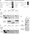
PCDH-γ interacts with PYK2 and FAK in vitro. A, a schematic of the domain structure of PCDH-γ protein. The shared cytoplasmic domain of PCDH-γ (γC; 125 amino acids) is marked. γC (amino acids 1–115) interacts with PYK2 and FAK in the yeast CytoTrap two-hybrid system. Cotransformation of the bait pSOS-γC with pMyr-PYK2 and pMyr-FAK into the cdc25H strain showed that both pMyr-PYK2 and pMyr-FAK interacted with pSOS-γC and rescued yeast growth defect at a restrictive temperature of 37 °C. pMyr-SB and pMyr-Lamin served as positive and negative controls, respectively. B, GST pull-down assay showed that immobilized GST-γC but not GST alone interacted with Myc-tagged PYK2 and FAK in 293T cell lysates. Shown are anti-Myc Western blots detecting PYK2 or FAK proteins (top two panels) and Coomassie staining of input GST proteins (middle). To demonstrate the direct interaction, purified His-PYK2 was incubated with GST-γC, and the associated His-PYK2 was detected by anti-PYK2 Western blot. C, PYK2 is associated with full-length PCDH-γ in 293T cells. Different combinations of PYK2 and PCDH-γ expression plasmids were transfected into 293T cells. Anti-FLAG immunoprecipitation was performed to isolate PYK2-associated protein complex (top) or PCDH-γ complex (bottom). The input and immunoprecipitated proteins were detected by Western blots. Full-length C5 PCDH-γ isoform was used in the experiments, and the interactions were verified in IP from both directions. D, FAK interacts with the C5 isoform of PCDH-γ in 293T cells. Similar experiments except for FAK were performed as in C.
FIGURE 2.
FIGURE 2.
PCDH-α interacts with PYK2 and FAK in vitro. A, PYK2 and FAK interact with αC in the GST pull-down assay. Like GST-γC, GST-αC interacted with Myc-tagged PYK2 or FAK from 293T cell lysates. B, the lysine-rich tail of γC but not that of αC is required for binding to FAK. GST pull-down assays using αC-(1–152), αC-(1–119), γC-(1–125), and γC-(1–115) demonstrated that the last 10 amino acids in γC are important for the binding of FAK to PCDH-γ but have less effect on the binding to PYK2. C and D, PYK2 and FAK interact with full-length PCDH-α in 293T cells. FLAG-tagged PYK2 or FAK proteins were expressed with full-length C2 isoform of PCDH-α in 293T cells. Anti-FLAG IP and Western blots show PYK2 (C) or FAK (D) is associated with PCDH-α.
FIGURE 3.
FIGURE 3.
PCDH-γ, PCDH-α, FAK, and/or PYK2 are in protein complexes in the brain. A, PCDH-α and PCDH-γ are associated with each other in 293T cells. Anti-FLAG IP showed that FLAG-tagged PCDH-α bound to PCDH-γ in cells transfected with both expression vectors. B and C, FAK and PYK2 interact with PCDH-γ-GFP fusion proteins in transfected 293T cells. D and E, PCDH-γ protein complexes consist of multiple PCDH proteins and FAK or PYK2. PCDH-γ containing protein complexes were affinity-purified with anti-GFP beads using Pcdh+/fusg brain tissues. The protein compositions of the complexes were analyzed by the indicated Western blots. P6 and P0 brain samples were used in D and E, respectively. F, PYK2 and FAK bind to the same C-terminal region in the shared cytoplasmic domain of PCDH-γ (amino acids 64–125). A GST pull-down assay using GSTγC-(1–71) and GSTγC-(64–125) is shown.
FIGURE 4.
FIGURE 4.
Mapping the interaction domains in FAK and PYK2 with PCDH-γ and PCDH-α. A, schematic drawings of domain organization of the wild type and deletion mutant PYK2 or FAK. The yeast growth at 37 °C indicates an interaction between different PYK2 and FAK protein with the bait pSOS-γC, demonstrating that the kinase domain of PYK2 interacts with γC. The relatively weak interaction of FAK with γC precluded further domain mapping. B and C, interactions between GSTγC and GSTαC with FAK (B) or PYK2 (C) deletion mutant proteins. The purified GSTγC and GSTαC were used to incubate with the cell lysates expressing different FAK or PYK2 deletion mutant proteins. GST pull-down assays show that γC interacts with the kinase domain in FAK (lane 7 in B) or PYK2 (lane 7, a weak band in C), and αC interacts with a region that contains both kinase and FAT domains (lane 14 in B and C). D, the cytoplasmic domains of PCDH-α and PCDH-γ are not substrates for FAK and PYK2 in vitro. In vitro kinase assays were carried out using purified recombinant FAK and PYK2 (100 ng/reaction). GST-γC and GST-αC (10 μg/reaction) were tested as their substrates. GST-N-Cad-C and GST alone served as positive and negative controls, respectively. Shown are the Coomassie staining of input substrate proteins and radioautography of [γ-32P]ATP kinase assays. N.D., not determined.
FIGURE 5.
FIGURE 5.
γC and αC inhibit FAK and PYK2 kinase activity in vitro. A and B, FLAG-tagged FAK (A) or PYK2 (B) were immunoprecipitated from 293T cell lysates. Identical aliquots of the purified kinases were assayed for their activity using E4Y1 (10 μg/reaction) as an exogenous substrate in the presence of increasing amounts of purified GST, GST-γC, and GST-αC, as indicated. The relative levels of E4Y1 phosphorylation as well as autophosphorylation of FAK or PYK2 were measured using a PhosphorImager. Relative kinase activity was normalized to kinase activity without any GST proteins. The means and S.E. of the relative kinase activities from three independent assays are shown. Data were analyzed using two-way analysis of variance (protein × dosage). p < 0.03. C, the effect of γC and αC on the kinase activity of FAK or PYK2 isolated from SYF cells. Shown are PhosphorImager acquired images of E4Y1 phosphorylation by FAK or PYK2. 5 μg of indicated GST fusion proteins were used in the kinase assay.
FIGURE 6.
FIGURE 6.
Cofractionation of PYK2 and FAK with PCDH. A, subcellular fractionation of brain lysates shows that FAK and PYK2 are present in the soluble cytosolic fraction (S2), membrane fractions (P2 and Syn), and PSD, whereas PCDH-γ is present in the membrane fraction and concentrated in the PSD. B, PCDH-γ-GFP proteins are overlapped with FAK and PYK2 in the dendrites of Pcdhfusg hippocampal neuron culture. Dendrites are identified by PSD95 staining. Bar, 20 μm. C, comparison of phosphorylation levels on Tyr(P)397 on FAK and Tyr(P)402 on PYK2 in the whole brain and total membrane samples indicates that membrane-bound FAK and PYK2 have lower kinase activity. Shown are panels of Western blots that compare levels of FAK, Tyr(P)397, PYK2, Tyr(P)402, PCDH-γ, and PCDH-α. Note that multiple bands of Tyr(P)402-PYK2 were detected in the membrane fractions that might reflect multiple phosphorylation events of PYK2. D, sucrose gradient ultracentrifugation analysis of total membrane proteins shows that Tyr(P)402-PYK2 is absent in the fractions that have significant overlap with PCDH proteins, whereas Tyr(P)397-FAK and FAK cofractionate. Shown are Western blots analyzing the abundance and distribution of indicated protein in 5–50% sucrose gradient fractions. The size markers for the sucrose gradient ultracentrifugation are bovine serum albumin (BSA; 66 kDa), thyroglobulin (THY; 669 kDa), and blue detran (BD; 2,000 kDa). E, the specificity of Tyr(P)402-PYK2 antibody is confirmed by Western blots of V5-tagged wild type PYK2 and Y402F PYK2 proteins overexpressed in 293T cells. pY, Tyr(P).
FIGURE 7.
FIGURE 7.
PCDH-γ and PCDH-α inhibit PYK2 and FAK activity in the brain. A and B, inactive PYK2 and FAK are associated with PCDH protein complex in the brain. Shown are comparisons of PYK2 and Tyr(P)402-PYK2 levels from Pcdh+/fusg total brain lysates and affinity-purified PCDH-γ protein complex with anti-GFP antibody (A). FAK and Tyr(P)397-FAK are shown in B. C, PYK2 is abnormally activated in Pcdhdel/del mice. Similar amounts of total brain membrane proteins from both wild type and Pcdhdel/del neonates were subjected to sucrose gradient ultracentrifugation and Western blot analyses. Each pair of Western blots from two genotypes was obtained with equal amounts of proteins in corresponded fractions and was probed and developed simultaneously. The abundance and distribution of PCDH-α, cadherins, and β-tubulin were almost identical, and they served as the internal controls for the experimental procedures. Tyr(P)402-PYK2 intensity was significantly up-regulated in the absence of PCDH-γ, whereas PYK2 protein level slightly decreased and the distribution remained the same in the mutants. Both FAK and Tyr(P)397-FAK levels were not significantly changed in Pcdhdel/del mice. pY, Tyr(P).
FIGURE 8.
FIGURE 8.
PYK2 overexpression induces apoptosis in the developing chicken spinal cord. A and B, overexpression PYK2 has no effect on cell proliferation in the chicken spinal cord. A, V5-tagged wild type and kinase-dead (KD) PYK2 were overexpressed in the right side of the spinal cord by electroporation at stage 12. Anti-phosphohistone H3 labeling (green cells on both sides of the central canal) shows that both electroporated (red) and nonelectroporated (dark) sides have similar numbers of mitotic cells 24 or 72 h after electroporation. B, BrdUrd administration to label S-phase proliferating cells shows that introduction of either wild type or kinase-dead PYK2 has little effect on BrdUrd incorporation. Note that in both A and B, V5-PYK2 is mostly localized in the neuronal processes, and phospho-H3 or BrdUrd labeling is exclusively in the nucleus. No apparent overlap of both signals was observed. C–E, overexpression of wild type but not kinase-dead PYK2 induces apoptosis in the spinal cord. C, representative images of anti-cleaved caspase-3 staining of chicken spinal cord sections that were electroporated with the indicated expression vectors. V5-tagged LacZ was used as a negative control. Embryos were harvested 24 h after electroporation. A small number of apoptotic cells were observed in the control sections due to the electroporation procedure. D, double-labeling of PYK2 (anti-V5; red) and active caspase-3 (green) shows that many caspase-3-positive nuclei were close to V5-stained soma or neuronal processes, suggesting that the apoptotic effect of PYK2 is probably cell-autonomous. E, quantitative analysis of the apoptotic cells in the PYK2 overexpressing spinal cord. Active caspase-3-positive cells were counted on sections from individual embryos. In the graph, the average number of active caspase-3-positive cells per section from individual embryos is plotted for each protein expressed. The results were subjected to t test and a p value of 0.024 was obtained between wild type and kinase-dead PYK2 samples. F, overexpression of PCDH-γ attenuates the apoptosis-inducing activity of PYK2. PCDH-γ and PYK2 expression vectors (at a ratio of 3:1) were electroporated into the chicken neural tubes as in C. A GFP expression vector was used as a negative control. The number of apoptotic cells was decreased upon co-expression of PYK2 with PCDH-γ, suggesting that PCDH-γ inhibits the activity of PYK2. The data were analyzed as in E. Bars,50 μmin A–C and 20 μmin D.

References

    1. Takeichi, M. (1995) Curr. Opin. Cell Biol. 7 619–627 - PubMed
    1. Yagi, T., and Takeichi, M. (2000) Genes Dev. 14 1169–1180 - PubMed
    1. Halbleib, J. M., and Nelson, W. J. (2006) Genes Dev. 20 3199–3214 - PubMed
    1. Wu, Q., and Maniatis, T. (1999) Cell 97 779–790 - PubMed
    1. Noonan, J. P., Grimwood, J., Schmutz, J., Dickson, M., and Myers, R. M. (2004) Genome Res. 14 354–366 - PMC - PubMed

Publication types

LinkOut - more resources