. 2008 Dec 1;68(23):9892-9.
doi: 10.1158/0008-5472.CAN-08-3134.

In vivo expansion, persistence, and function of peptide vaccine-induced CD8 T cells occur independently of CD4 T cells

Affiliations

In vivo expansion, persistence, and function of peptide vaccine-induced CD8 T cells occur independently of CD4 T cells

Deepak Assudani et al. Cancer Res. .

Abstract

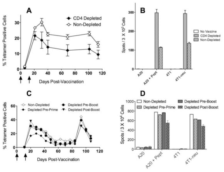
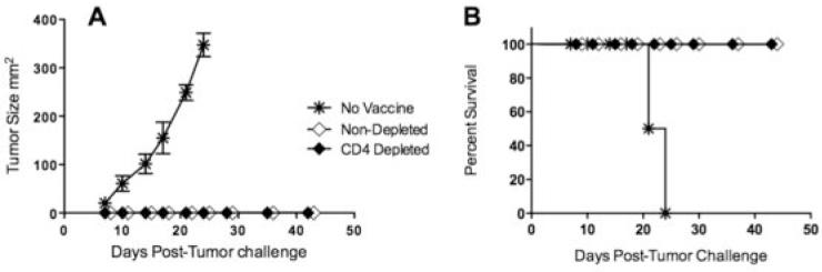
Significant efforts are being devoted toward the development of effective therapeutic vaccines against cancer. Specifically, well-characterized subunit vaccines, which are designed to generate antitumor cytotoxic CD8 T-cell responses. Because CD4 T cells participate at various stages of CD8 T-cell responses, it is important to study the role of CD4 T cells in the induction and persistence of antitumor CD8 T-cell responses by these vaccines. Recent evidence points to the requirement of CD4 T cells for the long-term persistence of memory CD8 T cells, which in the case of cancer immunotherapy would be critical for the prevention of tumor recurrences. The purpose of the present study was to assess whether CD4 T cells are necessary for the generation and maintenance of antigen-specific CD8 T cells induced by subunit (peptide or DNA) vaccines. We have used a vaccination strategy that combines synthetic peptides representing CD8 T-cell epitopes, a costimulatory anti-CD40 antibody and a Toll-like receptor agonist (TriVax) to generate large numbers of antigen-specific CD8 T-cell responses. Our results show that the rate of decline (clonal contraction) of the antigen-specific CD8 T cells and their functional state is not affected by the presence or absence of CD4 T cells throughout the immune response generated by TriVax. We believe that these results bear importance for the design of effective vaccination strategies against cancer.


Figures

Figure 1
Figure 1. TriVax immunization induces of large numbers of antigen-specific CD8 T cells
B6 mice (n = 3 per group) received 2 consecutive immunizations on days 0 and 21 (prime-boost) with TriVax/Poly-IC or TriVax/CpG prime-containing the Ova257-264 peptide. A, Antigen specific CD8 T cell responses were measured in peripheral blood by tetramer analysis at various time points. Dot plots for each animal are shown where the numbers in each plot represent the % tetramer positive cells (oval gates) of all CD8 positive cells. B, On day 84 (post prime), the in vivo antigen-specific effector cytolytic activity was estimated by injecting CFSE-labelled target cells and one day later the mice were euthanatized and tetramer positive cells were also measured in the spleens. Results obtained with naïve B6 mice show the absence of tetramer positive cells and persistence of Ova257-264-pulsed, CFSEhigh (arrow) target cells. On the other hand, all the vaccinated mice show tetramer positive spleen cells and almost complete absence of the peptide-pulsed CFSEhigh target cells. CFSElow target cells, which were not pulsed with peptide serve as an internal control were present in all the animals.
Figure 2
Figure 2. TriVax induced antigen specific CD8 T cells expand and persist in vivo in the absence of CD4 T cells
Untreated or continuously CD4 depleted B6 mice were primed either with Ova257-264-peptide TriVax (A) or ovalbumin DNA (B), followed 2 weeks later with a booster immunization with Ova257-264-peptide TriVax. CD4 depletion was initiated 3 days before the first vaccine administration and was continued throughout the entire experiment. Vertical arrows depict the vaccination time points. Antigen-specific CD8 T cell responses and CD4 T cell numbers in blood, were evaluated at various time points using Ova257-264-specific tetramers and anti-CD4 staining, respectively.
Figure 3
Figure 3. TriVax expanded antigen specific CD8 T cells exhibit effector activity in the absence of CD4 T cells
TriVax Ova257-264 immunized mice that were depleted or not of CD4 T cells were evaluated for the presence of effector CD8 T cells 3 months after the initial immunization. A, Mice from the experiment presented in Figure 2 were used for an in vivo cytotoxicity assay as described in Materials and Methods. Results obtained with naïve B6 mice (top panels) show persistence of Ova257-264-pulsed, CFSEhigh (arrows) target cells. On the other hand, the vaccinated mice show an almost complete absence of the peptide-pulsed CFSEhigh target cells. CFSElow target cells, which were not pulsed with peptide, serve as an internal control and were present in all the animals. B, IFN-γ ELISPOT assay using purified splenic CD8 T cells from Ova257-264 TriVax primed and boosted untreated and CD4-depleted mice against peptide pulsed tumor cells as well as ovalbumin-transfected tumors that naturally present the Ova257-264 epitope (EG7 and B16-Ova).
Figure 4
Figure 4. TriVax immunization in MHC-II-KO and CD4 T cell depleted mice yield similar results regarding the persistence of long-term CD8 T cell responses
Normal (untreated) B6, CD4 depleted and MHC-II-KO mice were primed with either TriVax Ova257-264 (A) or ovalbumin DNA (B) followed by a TriVax boost 3 weeks later. The vaccination time points are depicted by the vertical arrows. Peripheral blood lymphocytes were evaluated for the presence of antigen-specific CD8 T cells and CD4 positive periodically for a 3-month period.
Figure 5
Figure 5. DNA prime - TriVax boost CD8 T cell responses against an immunodominant tumor-reactive (p66) persist in vivo in the absence of CD4 T cells
BALB/c mice (CD4 T cell depleted or not) were primed with a DNA plasmid encoding a fragment of rNEU and were boosted 3 weeks later with p66 peptide TriVax. A, Antigen specific responses were monitored in blood using p66/Kd tetramer analysis periodically for over 3 months in untreated and in mice that were CD4 T depleted before the prime. B, Mice from the experiment described in panel A, were studied for their CD8 responses using an IFN-γ ELISPOT assay against peptide pulsed tumor cells (A20) as well as rNEU-transfected tumors (4T1-neu) that naturally present the p66 epitope. C, CD8 T cell responses from untreated or CD4-depleted mice (at either before the prime, before the boost or after the boost) were measured by tetramer analysis in blood samples. In addition, these mice received a second TriVax boost at day 83 to assess the capacity of these cells to re-expand for a second consecutive time. D, Mice from the experiment described in panel C were studied for their CD8 responses using an IFN-γ ELISPOT assay as described in panel B. Vertical arrows depict the vaccination time points.
Figure 6
Figure 6. Anti-tumor effector function of CD8 T cells expanded by TriVax immunization
Several groups of BALB/c mice (from Figs. 5A and 5B) were challenged subcutaneously with 3 × 10⁵ live TUBO breast cancer cells and monitored for tumor growth (A) and survival (B). A non-vaccinated group was added as control.

References

    1. Lanzavecchia A, Sallusto F. Understanding the generation and function of memory T cell subsets. Current Opin Immunol. 2005;17:326–32. - PubMed
    1. Prlic M, Williams MA, Bevan MJ. Requirements for CD8 T-cell priming, memory generation and maintenance. Current Opin Immunol. 2007;19:315–9. - PubMed
    1. Antia R, Ganusov VV, Ahmed R. The role of models in understanding CD8+ T-cell memory. Nat Rev Immunol. 2005;5:101–11. - PubMed
    1. Bennett SR, Carbone FR, Karamalis F, Flavell RA, Miller JF, Heath WR. Help for cytotoxic-T-cell responses is mediated by CD40 signalling. Nature. 1998;393:478–80. - PubMed
    1. Ridge JP, Di Rosa F, Matzinger P. A conditioned dendritic cell can be a temporal bridge between a CD4+ T-helper and a T-killer cell. Nature. 1998;393:474–8. - PubMed

Publication types

MeSH terms