. 2008 Dec 22;205(13):3119-31.
doi: 10.1084/jem.20082292. Epub 2008 Dec 1.

Yellow fever vaccine induces integrated multilineage and polyfunctional immune responses

Affiliations

Yellow fever vaccine induces integrated multilineage and polyfunctional immune responses

Denis Gaucher et al. J Exp Med. .

Abstract

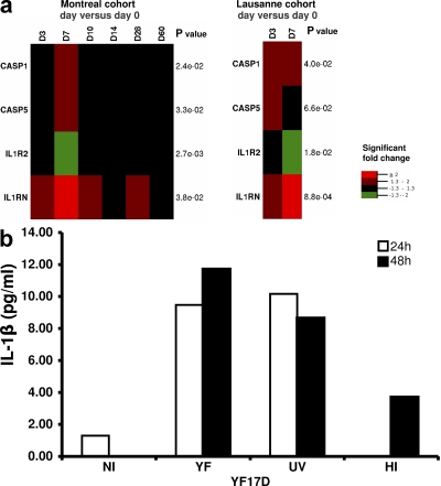
Correlates of immune-mediated protection to most viral and cancer vaccines are still unknown. This impedes the development of novel vaccines to incurable diseases such as HIV and cancer. In this study, we have used functional genomics and polychromatic flow cytometry to define the signature of the immune response to the yellow fever (YF) vaccine 17D (YF17D) in a cohort of 40 volunteers followed for up to 1 yr after vaccination. We show that immunization with YF17D leads to an integrated immune response that includes several effector arms of innate immunity, including complement, the inflammasome, and interferons, as well as adaptive immunity as shown by an early T cell response followed by a brisk and variable B cell response. Development of these responses is preceded, as demonstrated in three independent vaccination trials and in a novel in vitro system of primary immune responses (modular immune in vitro construct [MIMIC] system), by the coordinated up-regulation of transcripts for specific transcription factors, including STAT1, IRF7, and ETS2, which are upstream of the different effector arms of the immune response. These results clearly show that the immune response to a strong vaccine is preceded by coordinated induction of master transcription factors that lead to the development of a broad, polyfunctional, and persistent immune response that integrates all effector cells of the immune system.


Figures

Figure 1.
Figure 1.
Vaccination with YF17D induces early gene modulation. Global view of gene modulation in total blood cells after YF17D vaccination, as analyzed by gene array on the Montreal cohort (n = 9–15, depending on the time point). Heat map representation (a) and PCA (b) using the significantly modulated genes, in at least one comparison versus day 0.
Figure 2.
Figure 2.
Transcriptional network of differentially expressed genes after YF17D vaccination, as inferred by gene set enrichment. Network representation of inferred transcription factors (11) and predicted target genes that are significantly modulated. Node colors indicate fold change of gene expression between day 0 and 7 in n = 11 volunteers. Rectangular nodes indicate transcription factors identified by gene enrichment; the different shapes indicate genes in the different functional categories of Fig. 3 (a–e). Genes that were subsequently evaluated by RT-PCR are identified by an asterisk. IRF1, IRF8, and genes targeted by IRF1 or IRF8, but no other transcription factors in the figure were removed to increase readability (see Fig. S1 for a complete map). Fig. S1 is available at http://www.jem.org/cgi/content/full/jem.20082292/DC1.
Figure 3.
Figure 3.
YF17D vaccination stimulates multiple arms of the innate and adaptive immunity. (a–e) Heat map representation of significantly modulated genes, P values listed with each gene indicate the largest (least significant) P value among the significant fold changes. n = 9–15, depending on the time point. The listed genes fall under different functional categories: IFN-induced and TLR-associated genes (a); complement-associated genes (b); macrophage-associated genes (c); NK cell–associated genes (d); and B cell–associated genes (e). Supplemental document 2 includes a justification for the attribution of each gene into its respective group. Supplemental document 2 is available at http://www.jem.org/cgi/content/full/jem.20082292/DC1.
Figure 4.
Figure 4.
Heat map of fold change gene expression between day 0 and 7, as measured by qPCR. For a complete heat map of all measured fold changes see Fig. S3. Fig. S3 is available at http://www.jem.org/cgi/content/full/jem.20082292/DC1.
Figure 5.
Figure 5.
YF17D induces expression of genes associated with IL-1β and activates the inflammasome. (a) Heat maps showing the up-regulation of genes encoding inflammasome components and the modulation of other, IL-1β–associated genes. For IL-1R2 and -1RN only probes targeting all transcripts were considered. (b) IL-1β production by monocyte-derived DCs incubated with live (YF), UV-inactivated (UV), or heat-inactivated (HI) YF17D, as determined by ELISA. NI, noninfected cells. This represents the results from one representative of three separate experiments. The results for the two other experiments are shown in Fig. S4. Fig. S4 is available at http://www.jem.org/cgi/content/full/jem.20082292/DC1.
Figure 6.
Figure 6.
YF17D induces a mixed Th1/Th2 response. (a) Day 60 PBMCs were stimulated with the 22 peptide pools and assayed by CFSE labeling for their proliferative response. Bar graphs show data for six selected volunteers, and the dataset for all the volunteers can be found in Fig. S6 (c and d). At 24 h of culture, supernatants were analyzed by CBA to determine the Th1/Th2 cytokine secretion profile in response to each pool. The heat maps represent the data for the same six volunteers. The Th1/Th2 profiles determined this way for all the volunteers and pools are shown in Fig. S6 e. Fig. S6 is available at http://www.jem.org/cgi/content/full/jem.20082292/DC1.
Figure 7.
Figure 7.
YF17D vaccination induces specific CD4+ T cells of mixed T helper phenotype. (a) PBMCs from YF17D-vaccinated volunteers (day 28 after vaccination) were stimulated with a group of three immunostimulatory YF17D-derived peptides pools, and then stained with antibodies against CD4 (surface) and CD154, IL-2, and IFN-g (intracellular). The numbers indicate the percentages of cells within the parent population. (b) FACS analysis of PBMCs from day 365 after vaccination. Cells from four volunteers were stimulated (S) for with immunostimulatory YF17D peptide pools, and then restimulated (RS) or not with the same peptide pools before staining. The data were analyzed according to the gating strategy shown in Fig. S7, and are expressed as the percentage of central memory CD4+ T cells (CD45RA2CCR7+) that express the marker for recent activation CD154, and that are either Th1 or Th2 in the RS samples, over background (S). Fig. S7 is available at http://www.jem.org/cgi/content/full/jem.20082292/DC1.
Figure 8.
Figure 8.
YF17D elicits robust and diverse primary T helper cell responses in DC/T cell co-cultures. (a and b) Purified T helper cells were mixed with DCs that had been incubated with live or UV-inactivated YF17D. After 14 d, the cells were harvested and evaluated by flow cytometry for cytokine and CD154 expression after a short restimulation with autologous DCs targets that had been left untouched, pulsed with killed YF17D, or infected with live YF17D. All data plots show live CD4+-gated events. These are representative data of experiments done on cells from at least 10 volunteers. (c) Alternatively, the culture supernatants were harvested and evaluated by a multiplex cytokine analysis. Data representative of experiments done on cells from two volunteers.
Figure 9.
Figure 9.
Consensus transcriptional network of genes differentially expressed on day 7 as compared with day 0. Network representation of inferred transcription factors and predicted target genes that are consistently modulated in at least two out of three datasets, with the third dataset not being contradictory.

References

    1. Sekaly, R.P. 2008. The failed HIV Merck vaccine study: a step back or a launching point for future vaccine development? J. Exp. Med. 205:7–12. - PMC - PubMed
    1. Plotkin, S., and W. Orenstein. 2004. Vaccines. WB Saunders, Philadelphia.
    1. McMichael, A.J. 2006. HIV vaccines. Annu. Rev. Immunol. 24:227–255. - PubMed
    1. Monath, T.P. 2004. Yellow fever vaccine. In Vaccines. S. Plotkin and W. Orenstein, editors. WB Saunders, Philadelphia. 1095–1176.
    1. Bauer, J.H. 1931. The duration of passive immunity in yellow fever. Am. J. Trop. Med. Hyg. 11:451.

Publication types