. 2008 Dec 9;105(49):19183-7.
doi: 10.1073/pnas.0805514105. Epub 2008 Dec 1.

HDAC2 blockade by nitric oxide and histone deacetylase inhibitors reveals a common target in Duchenne muscular dystrophy treatment

Affiliations

HDAC2 blockade by nitric oxide and histone deacetylase inhibitors reveals a common target in Duchenne muscular dystrophy treatment

Claudia Colussi et al. Proc Natl Acad Sci U S A. .

Erratum in

  • Proc Natl Acad Sci U S A. 2009 Feb 3;106(5):1679. Steinkulher, Christian [corrected to Steinkuhler, Christian]

Abstract

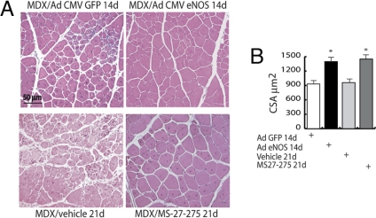
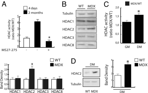
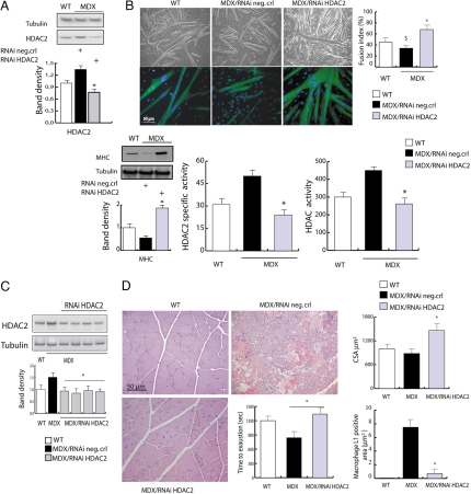
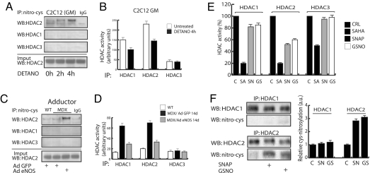
The overlapping histological and biochemical features underlying the beneficial effect of deacetylase inhibitors and NO donors in dystrophic muscles suggest an unanticipated molecular link among dystrophin, NO signaling, and the histone deacetylases (HDACs). Higher global deacetylase activity and selective increased expression of the class I histone deacetylase HDAC2 were detected in muscles of dystrophin-deficient MDX mice. In vitro and in vivo siRNA-mediated down-regulation of HDAC2 in dystrophic muscles was sufficient to replicate the morphological and functional benefits observed with deacetylase inhibitors and NO donors. We found that restoration of NO signaling in vivo, by adenoviral-mediated expression of a constitutively active endothelial NOS mutant in MDX muscles, and in vitro, by exposing MDX-derived satellite cells to NO donors, resulted in HDAC2 blockade by cysteine S-nitrosylation. These data reveal a special contribution of HDAC2 in the pathogenesis of Duchenne muscular dystrophy and indicate that HDAC2 inhibition by NO-dependent S-nitrosylation is important for the therapeutic response to NO donors in MDX mice. They also define a common target for independent pharmacological interventions in the treatment of Duchenne muscular dystrophy.


Conflict of interest statement

The authors declare no conflict of interest.

Figures

Fig. 1.
Fig. 1.
Adenovirus-mediated eNOS expression restores muscle differentiation in MDX mice. (A) H&E-stained sections of MDX adductor muscles injected with either GFP or eNOS adenovirus (1 × 10⁸ pfu per muscle) and evaluated 14 days after infection, or adductor muscles from MDX treated for 21 days with the class I HDAC inhibitor MS27-275 or the vehicle alone. (B) The bar graph shows the quantification of muscle fiber cross-sectional area (CSA) of the adductor from MDX mice injected with GFP or eNOS adenovirus and evaluated 14 days after infection, or adductor muscles from MDX treated for 21 days with the class I HDAC inhibitor MS27-275 or the vehicle alone. Cross-sectional area is expressed as mean ± SE (P < 0.05).
Fig. 2.
Fig. 2.
Class I HDAC expression and function is altered in MDX skeletal muscle and satellite cells. (A) Assessment of global HDAC activity was performed in whole lysates from WT and MDX mice adductor muscle by deacetylation assay. MS27-275 was added to fresh lysates at the concentration of 1 μM. Enzymatic activity is represented as the MDX/WT ratio. Experiments were performed 4 days and 2 months after birth (n = 6; P < 0.05). (B Upper) Western blotting analysis of class I HDACs in WT vs. MDX adductor skeletal muscle lysates. (B Lower) Data normalization by densitometric analysis. (C) Assessment of global HDAC activity was performed in lysates from primary satellite cells derived from single myofibers isolated from WT and MDX mice and cultured in growth medium (GM) or differentiation medium (DM). Enzymatic activity is represented as the MDX/WT ratio (n = 4; P < 0.05). (D) HDAC2 protein expression levels were measured by Western blot in primary satellite cells derived from single myofibers isolated from WT and MDX mice and cultured in DM. Data were normalized by densitometric analysis.
Fig. 3.
Fig. 3.
siRNA-mediated down-regulation of HDAC2 reproduces the effects of DIs and NO on MDX skeletal muscle. (A) Representative Western blotting analysis of HDAC2 levels in satellite cells from normal (WT) mice and MDX mice after delivery of scramble siRNA (control) or siRNA HDAC2. The densitometric analysis (Lower) shows that HDAC2 knockdown reduced the levels of endogenous HDAC2 to ≈50% of the endogenous one in MDX satellite cells. (B) The ability to differentiate into multinucleated myofibers was monitored in satellite cells from normal (WT) mice and MDX mice after delivery of scramble siRNA (control) or siRNA HDAC2. Shown are representative microphotographs of WT, MDX, and siRNA-treated MDX satellite cells at 72 h after differentiation. (B Upper) Phase contrast. (B Lower) MHC immunofluorescence and DAPI staining. Similar results were obtained with all of the oligonucleotides tested. Satellite cell differentiation has been evaluated by Western blotting analysis of the expression of MHC (Lower Left) and by the fusion index determined as the number of nuclei in myosin-expressing cells with >2 nuclei versus the total number of nuclei. Results are shown in the bar graph in Upper Right. HDAC global activity and HDAC2-specific activity have been evaluated in satellite cells treated with HDAC2-specific RNAi or control oligonucleotides and induced to differentiation. These results are shown in the lower, middle, and right panels of B. (C) Representative Western blotting analysis of HDAC2 levels in whole adductor muscles from normal (WT) mice vs. MDX mice treated with HDAC2 siRNAi or the scramble siRNA (control). Relative densitometric normalization was performed as described above (P < 0.05). (D) The in vivo HDAC2 knockdown shows morphological and functional recovery in MDX mice. The picture shows H&E staining of muscle sections and the treadmill test performed on WT and MDX mice injected with specific HDAC2 RNAi or scrambled oligonucleotides (neg.crl). The bar graph in Upper Right shows the average cross-section area in normal mice (white) and in MDX mice treated with HDAC2 RNAi (gray) or control oligonucleotides (black). The graph in Lower Right shows the presence of inflammatory infiltrate measured as macrophage L1-positive areas.
Fig. 4.
Fig. 4.
NO regulates the enzymatic activity of HDAC2 by cysteine-S-nitrosylation. (A) Western blotting analysis of HDAC1, HDAC2, and HDAC3 in C2C12 myoblasts after immunoprecipitation with anti S-nitroso-cysteine-specific antibody. An increased cysteine-S-nitrosylation of HDAC2 was detectable after 4 h of treatment with DETA-NO. (B) The bar graph shows the HDAC1-, HDAC2-, and HDAC3-specific activity in C2C12 evaluated in the presence or absence of DETA-NO treatment for 4 h. (C) Anti-S-nitroso-cysteine antibody immunoprecipitation and Western blotting analysis of HDAC1, HDAC2, and HDAC3 in WT and MDX mice infected with either eNOSS1177A or GFP adenovirus adductor muscles. An increased cysteine-S-nitrosylation of HDAC2 was detectable after 14 days from AdeNOS infection. (D) The bar graph shows in vivo the HDAC1-, HDAC2-, and HDAC3-associated enzymatic activity evaluated in adenovirus-infected WT and MDX mice expressing GFP or eNOS, respectively, in the adductor muscles. Assays were performed on total muscle lysates after immunoprecipitation with anti-HDAC1-, anti-HDAC2-, and anti-HDAC3-specific antibodies. (E) Evaluation of NO effect on recombinant E. coli GST-purified HDAC1-, HDAC2-, and HDAC3-specific activities in the presence or absence of HDAC inhibitor SAHA (SA, 5 μM) or NO donors SNAP (SN, 205 μM) or GSNO (GS, 250 μM). (F) Western blotting analysis of S-nitroso-cysteine on recombinant HDAC1 and HDAC2 proteins after NO donors SNAP (SN, 10 mM) or GSNO (GS, 10 mM). Data were normalized by densitometric analysis (Right).

References

    1. Brunelli S, et al. Nitric oxide release combined with nonsteroidal antiinflammatory activity prevents muscular dystrophy pathology and enhances stem cell therapy. Proc Natl Acad Sci USA. 2007;104:264–269. - PMC - PubMed
    1. Minetti GC, et al. Functional and morphological recovery of dystrophic muscles in mice treated with deacetylase inhibitors. Nat Med. 2006;12:1147–1150. - PubMed
    1. Iezzi S, et al. Deacetylase inhibitors increase muscle cell size by promoting myoblast recruitment and fusion through induction of follistatin. Dev Cell. 2004;6:673–684. - PubMed
    1. Pisconti A, et al. Follistatin induction by nitric oxide through cyclic GMP: A tightly regulated signaling pathway that controls myoblast fusion. J Cell Biol. 2006;172:233–244. - PMC - PubMed
    1. Bogdanovich S, et al. Functional improvement of dystrophic muscle by myostatin blockade. Nature. 2002;420:418–421. - PubMed

Publication types

MeSH terms

LinkOut - more resources