

Strong alkalinization in the anterior midgut of larval yellow fever mosquitoes (Aedes aegypti): involvement of luminal Na+/K+-ATPase
- PMID: 19048614
- PMCID: PMC2680433
- DOI: 10.1002/jez.512
Strong alkalinization in the anterior midgut of larval yellow fever mosquitoes (Aedes aegypti): involvement of luminal Na+/K+-ATPase
Abstract
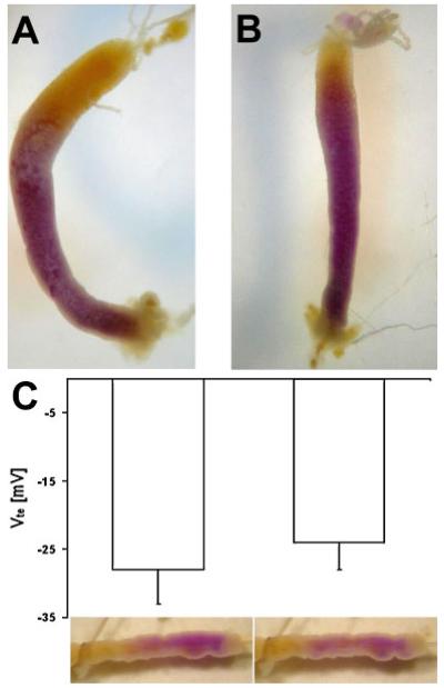
Recently, Na(+)/K(+)-ATPase has been detected in the luminal membrane of the anterior midgut of larval yellow fever mosquitoes (Aedes aegypti) with immunohistochemical techniques. In this study, the possible involvement of this ATPase in strong alkalinization was investigated on the level of whole larvae, isolated and perfused midgut preparations and on the molecular level of the Na(+)/K(+)-ATPase protein. Ouabain (5 mM) did not inhibit the capability of intact larval mosquitoes to alkalinize their anterior midgut. Also in isolated and perfused midgut preparations the perfusion of the lumen with ouabain (5 mM) did not result in a significant change of the transepithelial voltage or the capacity of luminal alkalinization. Na(+)/K(+)-ATPase activity was completely abolished when KCl was substituted with choline chloride, suggesting that the enzyme cannot act as an ATP-driven Na(+)/H(+)-exchanger. Altogether the results of the present investigation indicate that apical Na(+)/K(+)-ATPase is not of direct importance for strong luminal alkalinization in the anterior midgut of larval yellow fever mosquitoes.
Figures
References
-
- Bernick EP, Onken H, Kirschner LB, Moffett DF. ATPase activities in Aedes aegypti midgut; SICB 2004 Annual Meeting; 2004. p. 345p. Abstracts, 2004.
-
- Bijelic G, O'Donnell MJ. Diuretic factors and second messengers stimulate secretion of the organic cation TEA by the Malpighian tubules of Drosophila melanogaster. J Insect Physiol. 2005;51:267–275. - PubMed
-
- Boudko DY, Moroz LL, Linser PJ, Trimarchi JR, Smith PJS, Harvey WR. In situ analysis of pH gradients in mosquito larvae using non-invasive, self-referencing, pH-sensitive microelectrodes. J Exp Biol. 2001b;204:691–699. - PubMed
-
- Bradford MM. A rapid and sensitive method for the quantitation of microgram quantities of protein utilizing the principle of protein–dye binding. Anal Biochem. 1976;72:248–254. - PubMed
Publication types
MeSH terms
Substances
Grants and funding
LinkOut - more resources
Full Text Sources
