. 2009 Apr;24(4):597-605.
doi: 10.1359/jbmr.081210.

Osteocyte apoptosis controls activation of intracortical resorption in response to bone fatigue

Affiliations

Osteocyte apoptosis controls activation of intracortical resorption in response to bone fatigue

Luis Cardoso et al. J Bone Miner Res. 2009 Apr.

Abstract

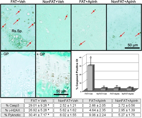
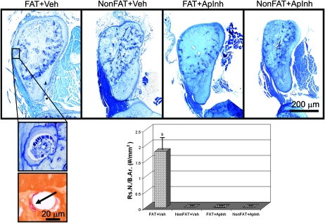
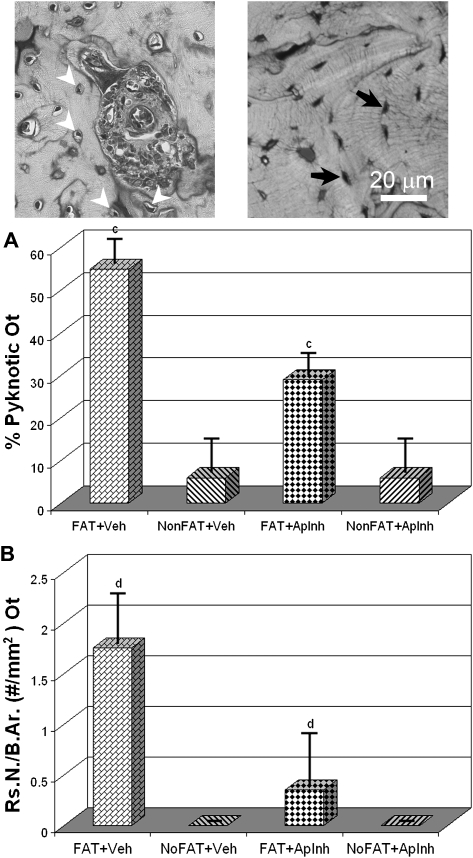
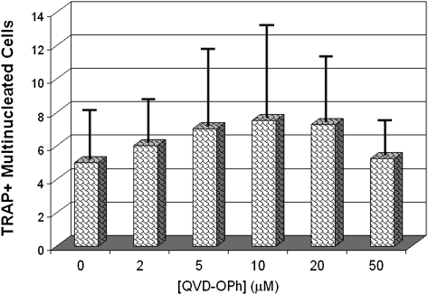
Osteocyte apoptosis is spatially and temporally linked to bone fatigue-induced microdamage and to subsequent intracortical remodeling. Specifically, osteocytes surrounding fatigue microcracks in bone undergo apoptosis, and those regions containing apoptotic osteocytes co-localize exactly with areas subsequently resorbed by osteoclasts. Here we tested the hypothesis that osteocyte apoptosis is a key controlling step in the activation and/or targeting of osteoclastic resorption after bone fatigue. We carried out in vivo fatigue loading of ulna from 4- to 5-mo-old Sprague-Dawley rats treated with an apoptosis inhibitor (the pan-caspase inhibitor Q-VD-OPh) or with vehicle. Intracortical bone remodeling and osteocyte apoptosis were quantitatively assessed by standard histomorphometric techniques on day 14 after fatigue. Continuous exposure to Q-VD-OPh completely blocked both fatigue-induced apoptosis and the activation of osteoclastic resorption, whereas short-term caspase inhibition during only the first 2 days after fatigue resulted in >50% reductions in both osteocyte apoptosis and bone resorption. These results (1) show that osteocyte apoptosis is necessary to initiate intracortical bone remodeling in response to fatigue microdamage, (2) indicate a possible dose-response relationship between the two processes, and (3) suggest that early apoptotic events after fatigue-induced microdamage may play a substantial role in determining the subsequent course of tissue remodeling.


Figures

FIG. 1
FIG. 1
Continuous Q-VD-OPh completely prevented fatigue-induced increases in osteocyte apoptosis. Photomicrographs show immunostaining for cleaved capsase-3. (Top panels) Osteocyte staining in fatigue-loaded and nonfatigued ulnar cortical bone, both with and without apoptosis inhibitor. (Bottom panels) Negative (L) and positive (R) tissue controls in growth plates with strong staining of hypertrophic chondrocytes. Large numbers of caspase positive-stained (Casp+) apoptotic osteocytes (arrows pointing to brown stained cells) are evident in ulnar bone of fatigue-loaded, vehicle-treated (FAT + Veh) animals. In contrast, Casp+ osteocytes in bones from fatigued animals treated with the apoptosis inhibitor (FAT + ApInh) were comparable to nonfatigued controls. Apoptotic osteocyte counts are summarized in the accompanying graph for Casp+ cells and in the table for other apoptosis assays (H2AX and pyknotic cells, a p < 0.001 vs. nonfatigued controls). Results were similar for all apoptosis markers.
FIG. 2
FIG. 2
Continuous apoptosis suppression with Q-VD-OPh completely prevented fatigue-induced activation of intracortical remodeling. Photomicrographs show cross-sections of ulnar mid-diaphyses from fatigue-loaded and nonfatigued control ulnar cortical bone, both with and without apoptosis inhibitor treatment. Intracortical remodeling spaces were present in FAT+Veh bone but were not observed in bones from fatigued animals treated with the apoptosis inhibitor (FAT + ApInh); enlargements below show a toluidine blue–stained section (top) through the infilling region of a remodeling space (enlarged from low power image above) and a TRACP-stained section (bottom) through the cutting cone from a different level of the remodeling space (arrow = red TRACP+-stained osteoclast). Histomorphometry data for resorption space number (Rs.N/B.Ar, #/mm2 bone area) show a complete suppression of fatigue-induced activation of intracortical resorption (b p < 0.0001 vs. nonfatigue controls).
FIG. 3
FIG. 3
Effects of short-duration apoptosis inhibition on osteocyte apoptosis and intracortical resorption at 14 days after loading. Treatment with Q-VD-OPh for 2 days immediately after fatigue loading resulted in a nearly 50% reduction of osteocyte apoptosis compared with vehicle treated animals. Data are shown for pyknotic osteocytes (A; c p < 0.01); the accompanying photomicrographs show pyknotic, retracted osteocytes (left photo, white arrows) and normal osteocytes in control bone (right photo, black arrows). Acute treatment with apoptosis inhibitor suppressed the fatigue-induced activation of intracortical resorption by ∼75% relative to vehicle-treated animals (B, d p < 0.025).
FIG. 4
FIG. 4
Q-VD-OPh effect on in vitro osteoclast differentiation in total rat bone marrow cultures. Numbers of TRACP+ multinucleated cells were unaffected by the apoptosis inhibitor. Thus, the ability of Q-VD-OPh to prevent bone resorption in vivo does not seem to result from direct inhibition of osteoclastogenesis.

Similar articles

Cited by

References

    1. Burr DB, Martin RB, Schaffler MB, Radin EL. Bone remodeling in response to in vivo fatigue microdamage. J Biomech. 1985;18:189–200. - PubMed
    1. Enlow DH. Functions of the Haversian system. Am J Anat. 1962;110:269–305. - PubMed
    1. Parfitt AM. Bone age, mineral density, and fatigue damage. Calcif Tissue Int. 1993;53(Suppl 1):S82–S85. - PubMed
    1. Schaffler MB. Role of bone turnover in microdamage. Osteoporos Int. 2003;14(Suppl 5):73–80. - PubMed
    1. Bentolila V, Boyce TM, Fyhrie DP, Drumb R, Skerry TM, Schaffler MB. Intracortical remodeling in adult rat long bones after fatigue loading. Bone. 1998;23:275–281. - PubMed

Publication types

MeSH terms