. 2008 Dec 23;47(51):13604-9.
doi: 10.1021/bi801884z.

Alpha-Synuclein conformation affects its tyrosine-dependent oxidative aggregation

Affiliations

Alpha-Synuclein conformation affects its tyrosine-dependent oxidative aggregation

Rebecca A S Ruf et al. Biochemistry. .

Abstract

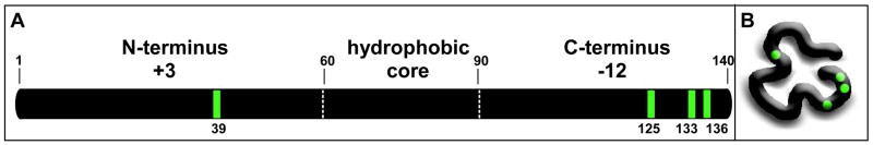
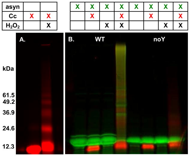
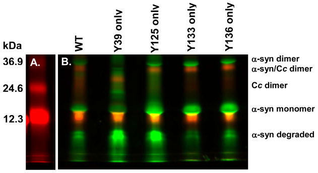
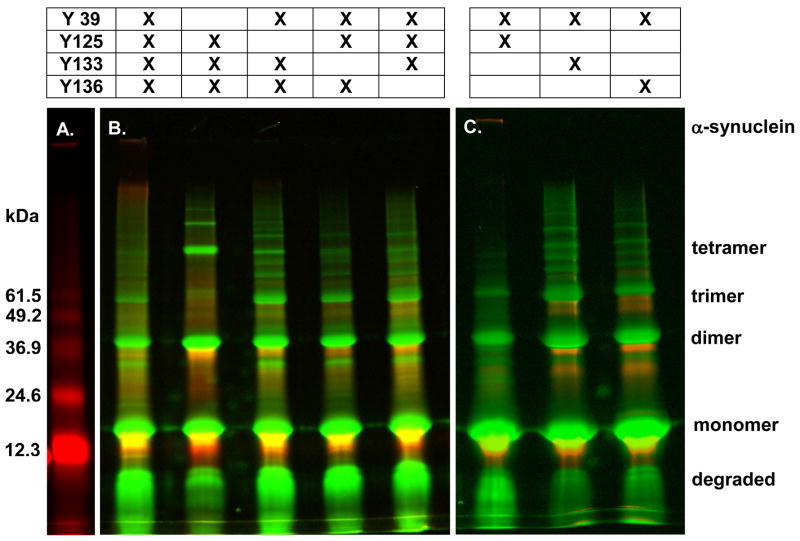
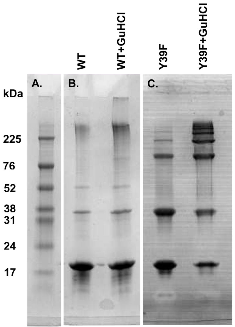
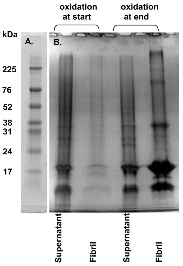
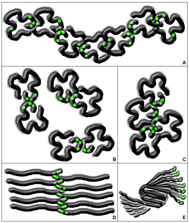
Oxidative stress and aggregation of the protein alpha-synuclein are thought to be key factors in Parkinson's disease. Previous work shows that cytochrome c with H(2)O(2) causes tyrosine-dependent in vitro peroxidative aggregation of proteins, including alpha-synuclein. Here, we examine the role of each of alpha-synuclein's four tyrosine residues and how the protein's conformation affects covalent oxidative aggregation. When alpha-synuclein adopts a collapsed conformation, tyrosine 39 is essential for wild-type-like covalent aggregation. This lone N-terminal tyrosine, however, is not required for wild-type-like covalent aggregation in the presence of a denaturant or when alpha-synuclein is present in noncovalent fibrils. We also show that preformed oxidative aggregates are not incorporated into noncovalent fibrils. These data provide insight into how dityrosine may be formed in Lewy bodies seen in Parkinson's disease.


Figures

Figure 1
Figure 1
∝-Synuclein structure and conformation. A schematic representation of α-synuclein’s primary structure showing relevant regions, net charges, and position numbers (A), and a cartoon of its collapsed conformation (B). The positions of the tyrosine residues are indicated in green.
Figure 2
Figure 2
Alexa Fluor labeling detects peroxidative aggregation. (A) Cytochrome c was incubated with and without H2O2. (B) ∝-Synuclein, either the wild-type protein with the Alexa Fluor label at position 3 (WT, lanes 1–4), or a no tyrosine variant with the Alexa Fluor label at position 3 (noY, lanes 5–8) were combined with various combinations of cytochrome c and H2O2 for 90 min., separated on a 10–20% gradient polyacrylamide gel, and visualized by fluorescence (green, α-synuclein; red, cytochrome c).
Figure 3
Figure 3
Every tyrosine in α-synuclein is reactive, but to varying extents. (A) Cytochrome c was incubated with H2O2. (B) The wild-type protein (WT) and single tyrosine-containing variants of α-synuclein were reacted with cytochrome c and H2O2 for 90 minutes, and treated as described in the caption to Figure 2.
Figure 4
Figure 4
At least one tyrosine on each end is required for wild-type-like covalent aggregation of collapsed α-synuclein. (A) Cytochrome c was incubated with H2O2. (B) The wild-type protein and variants containing three tyrosines or (C) two tyrosines were reacted with cytochrome c and H2O2 for 90 minutes, and treated as described in the caption to Figure 2. Legend at top indicates which tyrosines are present in each variant. α-Synuclein aggregate species are indicated on the right.
Figure 5
Figure 5
Tyrosine 39 is not required for full aggregation of denatured α-synuclein. (A) Molecular weight marker. (B) The wild-type protein (WT) and (C) the variant with tyrosine 39 removed (Y39F) were reacted with cytochrome c and H2O2 in the presence (lanes 2) or absence (lanes 1) of guanidine hydrochloride. Samples were separated on a 10–20% gradient polyacrylamide gel, and visualized by Coomassie Blue staining.
Figure 6
Figure 6
Oxidative aggregation interferes with fibril formation. (A) Molecular weight marker. (B) The wild-type protein was reacted with cytochrome c and H2O2 before (lanes 1 and 2) or after (lanes 3 and 4) fibril formation. Fibrils were isolated by centrifugation. Fibrils (lanes 2 and 4) and the supernatant (lanes 1 and 3) were boiled with SDS, separated on 10–20% gradient polyacrylamide gels, and visualized with Coomassie Blue.
Figure 7
Figure 7
α-Synuclein conformation and covalent aggregation. When α-synuclein adopts a collapsed conformation, various covalently aggregated species are more equally populated when at least one tyrosine is present on each end of the protein (A). Even-numbered aggregates are favored when tyrosine 39 is removed (B and C). When α-synuclein is completely disordered, the largest aggregate species are favored, even if tyrosine 39 is removed (D). Covalent α-synuclein aggregates are unable to fold into the beta-sheet aggregation nucleus, but once the folded monomers have assembled into protofibrils, the termini are stacked, allowing covalent aggregation of any of the tyrosines (E).

References

    1. Hague SM, Klaffke S, Bandmann O. Neurodegenerative disorders: Parkinson’s disease and Huntington’s disease. J Neurol Neurosurg Psychiatr. 2005;76:1058–1063. - PMC - PubMed
    1. National Parkinson’s foundation.

    1. Spillantini MG, Schmidt ML, Lee VMY, Trojanowski JQ, Jakes R, Goedert M. Alpha-synuclein in Lewy bodies. Nature. 1997;388:839–840. - PubMed
    1. Hashimoto M, Takeda A, Hsu LJ, Takenouchi T, Masliah E. Role of cytochrome c as a stimulator of alpha-synuclein aggregation in Lewy body disease. J Biol Chem. 1999;274:28849–28852. - PubMed
    1. D’Andrea MR, Ilyin S, Plata-Salaman CR. Abnormal patterns of microtubule-associated protein-2 (map-2) immunolabeling in neuronal nuclei and Lewy bodies in Parkinson’s disease substantia nigra brain tissues. Neuroscience Letters. 2001;306:137–140. - PubMed

Publication types