. 2009 Jan 30;284(5):3345-3353.
doi: 10.1074/jbc.M808396200. Epub 2008 Dec 2.

Transglutaminase-1 regulates renal epithelial cell proliferation through activation of Stat-3

Affiliations

Transglutaminase-1 regulates renal epithelial cell proliferation through activation of Stat-3

Zhu Zhang et al. J Biol Chem. .

Abstract

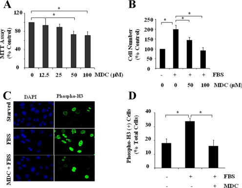
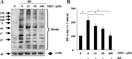
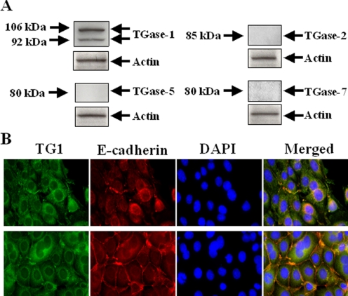
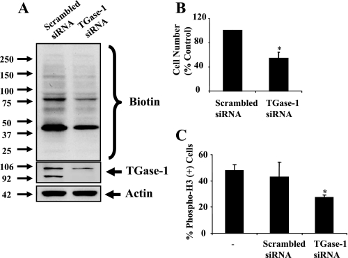
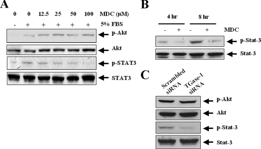
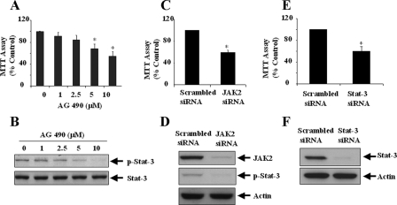
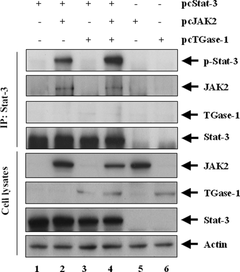
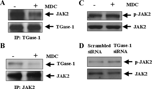
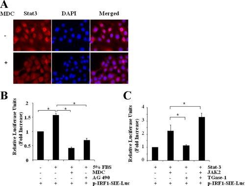
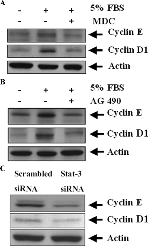
Transglutaminase-1 (TGase-1) is a Ca(2+)-dependent enzyme capable of cross-linking a variety of proteins and promoting wound healing in the skin. In this study, we examined the role of TGase-1 in proliferation of renal proximal tubular cells (RPTC). TGase-1, but not TGase-2, -5, and -7, was expressed in RPTC. Treatment with monodansylcadarevine (MDC), a selective TGase inhibitor or down-regulation of TGase-1 with small interfering RNA (siRNA) decreased RPTC proliferation. Proliferation of RPTC was accompanied by activation of Akt and Stat-3 (signal transducer and activator of transcription-3). Treatment with MDC or TGase-1 siRNA decreased Stat-3 but not Akt phosphorylation. Further studies showed that the Janus-activated kinase 2 (JAK2) mediates phosphorylation of Stat-3, and knockdown of either JAK2 or Stat-3 by siRNA decreased RPTC proliferation. However, inhibition of TGase-1 decreased phosphorylation of Stat-3 but not JAK2. Overexpression of Stat-3, JAK2, and/or TGase-1 in RPTC revealed that JAK2 is indispensable for TGase-1 to induce Stat-3 phosphorylation and TGase-1 potentiates JAK2-induced Stat-3 phosphorylation. Consistent with these observations, we found that inhibition of TGase-1 and the JAK2-Stat-3 signaling pathway decreased the transcriptional activity of Stat-3 and expression of the Stat-3-targeted genes, cyclin D1 and cyclin E. Conversely, overexpresssion of TGase-1 enhanced the JAK2-dependent transcriptional activity of Stat-3. Finally, TGase-1 was found to interact with JAK2, and this interaction was inhibited by MDC. These results demonstrate that TGase-1 plays an important role in regulation of renal epithelial cell proliferation through the JAK2-Stat-3 signaling pathway.


Figures

FIGURE 1.
FIGURE 1.
Concentration-dependent inhibition of cell proliferation by MDC in cultured RPTC. RPTC were cultured for 24 h in the presence of 5% FBS and then treated with MDC for an additional 48 h at the indicated concentrations (A and B). Cell proliferation was assessed by the MTT assay (A) or by counting cell numbers (B). RPTC were starved for 24 h and then incubated in the presence of 5% FBS with 100 μm MDC for an additional 48 h (C and D). Cells were stained with DAPI and anti-phospho-H3 antibody, and photographs were taken (×600 magnification) (C). Cell proliferation was evaluated by percentage of cells with phospho-H3-positive nuclei. D, data are means ± S.D. of three independent experiments conducted in triplicate and expressed as the percentage of control or total cell number. *, p < 0.05.
FIGURE 2.
FIGURE 2.
Effect of MDC on serum-induced TGase activity in cultured RPTC. RPTC were cultured in the medium containing 5% FBS in the absence or presence of MDC for 24 h at the indicated concentrations, and then 1 mm (5-(biotinamido)pentylamine) BP was added to the culture and incubated for an additional 1 h. Cells were harvested for analysis of TGase transamidation activity, as described under “Materials and Methods.” A representative blot from three independent experiments is shown (A). The biotin-labeled proteins were quantified by densitometry and normalized to actin B, data are means ± S.D. of three independent experiments and expressed as percentage of expression level relative to control. *, p < 0.05.
FIGURE 3.
FIGURE 3.
Expression of TGases in RPTC. RPTC were cultured to 60-70% confluence. Then cell lysates were prepared and subjected to immunoblot analysis using antibodies to TGase-1, TGase-2, TGase-5, TGase-7, or actin (A). RPTC were fixed and then stained using antibodies against TGase-1 and E-cadherin followed by Alexa Fluor 488-conjugated anti-rabbit IgG, or Texas Red goat anti-rabbit IgG and DAPI (B). Representative photographs were taken from a nonconfluent (upper row) and a confluent area (lower row).
FIGURE 4.
FIGURE 4.
Effect of siRNA specific to TGase-1 on RPTC proliferation. RPTC were transfected with scrambled siRNA or siRNA specific to TGase-1 and then cultured for 48 h in DMEM with 0.5% FBS. A, cells were stimulated with 5% FBS for 1 h in the presence of 1 mm BP. TG transamidation activity was analyzed as described in the legend to Fig. 2. B, cells were incubated for an additional 48 h in the presence of 5% FBS, and cell number was counted. C, cells were incubated for an additional 48 h in the presence of 5% FBS and cell proliferation was evaluated by counting cells with phospho-H3 staining. Data are means ± S.D. of three independent experiments conducted in triplicate and expressed as the percentage of cells treated with scrambled siRNA or total cell number. *, p < 0.05.
FIGURE 5.
FIGURE 5.
Effect of MDC on phosphorylation of Akt, and Stat-3 in RPTC. A, RPTC were serum-starved for 24 h and then stimulated with 5% FBS for 1 h (A) or 4 and 8 h (B) in the presence or absence of 100 μm MDC. Cell lysates were analyzed by immunoblot analysis with antibodies against phospho-Akt, Akt, phospho-Stat-3, or Stat-3. C, RPTC were transfected with scrambled siRNA or siRNA specific to TGase-1 and then cultured for an additional 48 h in the presence of 0.5% FBS. After stimulation with 5% FBS for 1 h, cell lysates were analyzed by immunoblot analysis with antibodies against phospho-Akt, Akt, phospho-Stat-3, or Stat-3. Representative immunoblots from three or more experiments are shown.
FIGURE 6.
FIGURE 6.
Effect of the JAK/Stat-3 pathway inhibition on RPTC proliferation. RPTC were cultured for 24 h in the presence of 5% FBS and then treated with AG490 at the indicated concentrations for an additional 24 h (A) or 1 h (B). A, cell proliferation was assessed by the MTT assay. Data are means ± S.D. *, p < 0.05, compared with control group. B, cell lysates were subjected to immunoblot analysis with antibodies against phospho-Stat-3 and Stat-3. Representative immunoblots from three or more experiments are shown. RPTC were transfected with scrambled siRNA or siRNA specific to JAK2 or Stat-3 and then cultured for 48 h in DMEM with 0.5% FBS (C-F). Cells were incubated for an additional 48 h in the presence of 5% FBS, and cell proliferation was evaluated by the MTT assay (C and E). Cells were stimulated with 5% FBS for 1 h, and cell lysates were analyzed by immunoblot analysis with antibodies against phospho-Stat-3, JAK2 (D), Stat-3, or actin (F).
FIGURE 7.
FIGURE 7.
TGase-1 coexpression enhances JAK2-dependent phosphorylation of Stat-3. RPTC were transiently transfected with plasmids encoding wild types of TGase-1, JAK2, or Stat-3, either alone or in different combinations as indicated. After 48 h, the cells were harvested and Stat-3 was immunoprecipitated (IP) with an anti-Stat-3 antibody. Immunoprecipitates and cell lysates were analyzed by immunoblotting with the indicated antibodies. Representative immunoblots from three experiments are shown.
FIGURE 8.
FIGURE 8.
Inhibition of TGase-1 disrupts the interaction of TGase-1 with JAK2 but does not affect serum-induced JAK2 phosphorylation. A and B, RPTC were transiently transfected with plasmids encoding TGase-1 and JAK2. TGase-1 was immunoprecipitated (IP) with an anti-TGase-1 antibody and then analyzed with antibodies against JAK2 or TGase-1 (A). JAK2 was immunoprecipitated with an anti-JAK2 antibody and then analyzed with antibodies against TGase-1 or JAK2 (B). RPTC were treated with MDC (100 μm) for 1 h (C) or TGase-1 siRNA for 48 h (D) and then harvested for immunoblot analysis with antibodies to phospho-JAK2 or JAK2. Representative immunoblots from three experiments are shown.
FIGURE 9.
FIGURE 9.
The role of TGase-1 in serum-stimulated nuclear accumulation of Stat-3 and Stat-3 transcriptional activity. A, RPTC were cultured for 24 h in DMEM/F-12 with 5% FBS in the presence or absence of 100 μm MDC and then stained with anti-Stat-3 antibody and DAPI. B, RPTC were transfected with p-IRF1-SIE-Luc and serum-starved for 24 h and then incubated with DMEM alone, DMEM plus 5% FBS, or DMEM plus 5% FBS containing 100 μm MDC or 10 μm AG490 for an additional 24 h. C, RPTC were transfected with plasmids encoding wild types of TGase-1, JAK2, or Stat-3, either alone or in different combinations as indicated and then incubated for 24 h with DMEM plus 5% FBS. Cells were lysed, and equal amounts of protein were used to evaluate the luciferase activity by dual luciferase reporter assay as described under “Materials and Methods.” Data are means ± S.E. of three independent experiments conducted in triplicate and expressed as -fold increase relative to controls. *, p < 0.05.
FIGURE 10.
FIGURE 10.
Effect of MDC, AG490, and Stat-3 siRNA on cyclin D1 and cyclin E expression. A and B, RPTC were serum-starved for 24 h and then incubated with 5% FBS for an additional 24 h in the presence or absence of 100 μm MDC (A) or 10 μm AG490 (B). C, RPTC were transfected with scrambled siRNA or siRNA specific to TGase-1 and then cultured for 48 h in DMEM with 5% FBS. Cell lysates were subjected to immunoblot analysis with antibodies against cyclin D1 and cyclin E. Representative immunoblots from three or more experiments are shown.

References

    1. Lorand, L., and Graham, R. M. (2003) Nat. Rev. Mol. Cell. Biol. 4, 140-156 - PubMed
    1. Inada, R., Matsuki, M., Yamada, K., Morishima, Y., Shen, S. C., Kuramoto, N., Yasuno, H., Takahashi, K., Miyachi, Y., and Yamanishi, K. (2000) Am. J. Pathol. 157, 1875-1882 - PMC - PubMed
    1. Hiiragi, T., Sasaki, H., Nagafuchi, A., Sabe, H., Shen, S. C., Matsuki, M., Yamanishi, K., and Tsukita, S. (1999) J. Biol. Chem. 274, 34148-34154 - PubMed
    1. Martinet, N., Bonnard, L., Regnault, V., Picard, E., Burke, L., Siat, J., Grosdidier, G., Martinet, Y., and Vignaud, J. M. (2003) Am. J. Respir. Cell Mol. Biol. 28, 428-435 - PubMed
    1. Fesus, L., and Piacentini, M. (2002) Trends Biochem. Sci. 27, 534-539 - PubMed

Publication types