. 2009 Feb 20;284(8):5352-61.
doi: 10.1074/jbc.M805768200. Epub 2008 Dec 1.

The endoplasmic reticulum enzyme DGAT2 is found in mitochondria-associated membranes and has a mitochondrial targeting signal that promotes its association with mitochondria

Affiliations

The endoplasmic reticulum enzyme DGAT2 is found in mitochondria-associated membranes and has a mitochondrial targeting signal that promotes its association with mitochondria

Scot J Stone et al. J Biol Chem. .

Abstract

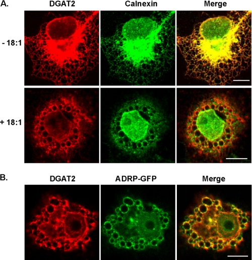
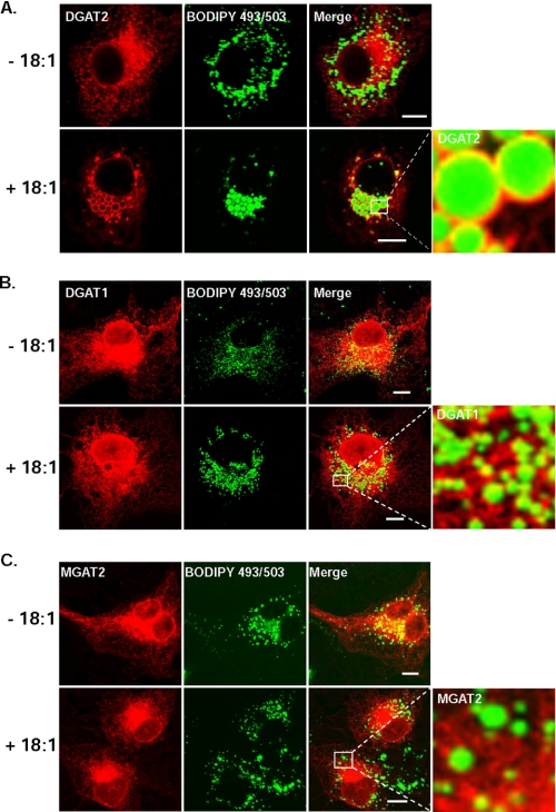
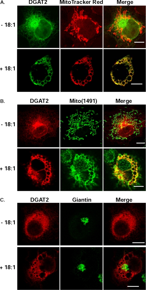
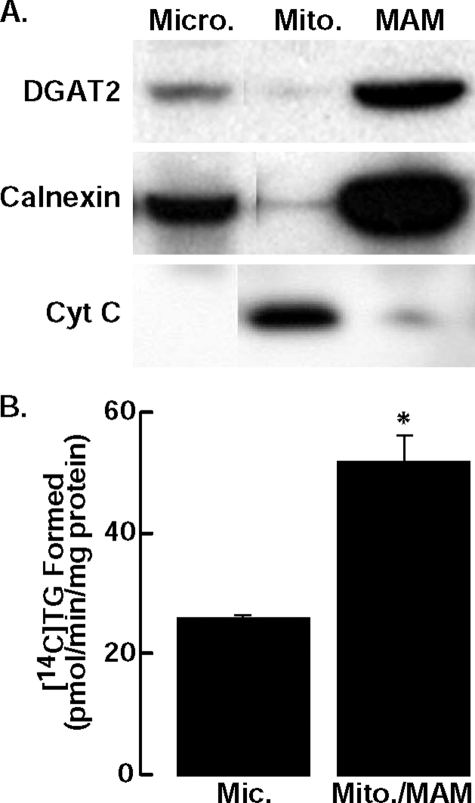
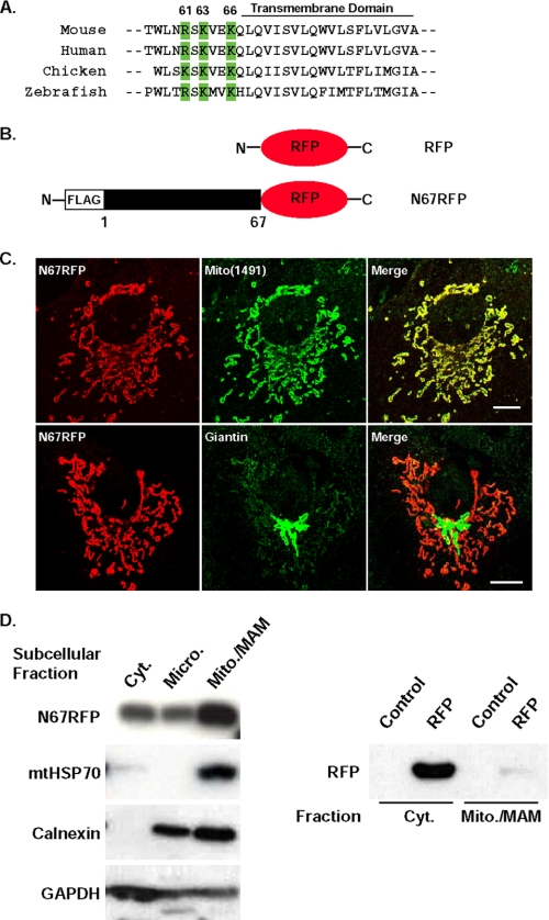
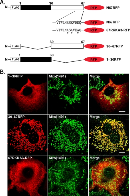
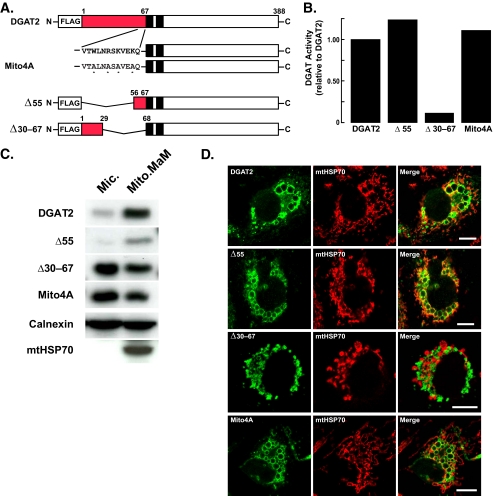
The synthesis and storage of neutral lipids in lipid droplets is a fundamental property of eukaryotic cells, but the spatial organization of this process is poorly understood. Here we examined the intracellular localization of acyl-CoA:diacylglycerol acyltransferase 2 (DGAT2), an enzyme that catalyzes the final step of triacylglycerol (TG) synthesis in eukaryotes. We found that DGAT2 expressed in cultured cells localizes to the endoplasmic reticulum (ER) under basal conditions. After providing oleate to drive TG synthesis, DGAT2 also localized to near the surface of lipid droplets, where it co-localized with mitochondria. Biochemical fractionation revealed that DGAT2 is present in mitochondria-associated membranes, specialized domains of the ER that are highly enriched in lipid synthetic enzymes and interact tightly with mitochondria. The interaction of DGAT2 with mitochondria depended on 67 N-terminal amino acids of DGAT2, which are not conserved in family members that have different catalytic functions. This targeting signal was sufficient to localize a red fluorescent protein to mitochondria. A highly conserved, positively charged, putative mitochondrial targeting signal was identified in murine DGAT2 between amino acids 61 and 66. Thus, DGAT2, an ER-resident transmembrane domain-containing enzyme, is also found in mitochondria-associated membranes, where its N terminus may promote its association with mitochondria.


Figures

FIGURE 1.
FIGURE 1.
Oleate loading alters the subcellular distribution of DGAT2. Confocal immunofluorescence microscopy of DGAT2 transiently expressed in COS-7 cells. A, COS-7 cells transfected with DGAT2 were treated with 0.5 mm oleate (+18:1) for 12 h or left untreated (-18:1) and then stained with anti-FLAG and anti-calnexin antibodies. (The nuclear calnexin staining (lower panel) likely reflects nuclear membrane staining in an image from a plane that did not bisect the nucleus.) B, DGAT2 co-localizes with ADRP-GFP. COS-7 cells were co-transfected with DGAT2 and ADRP-GFP. Transfected cells were then incubated with 0.5 mm oleate for 12 h. Cells were fixed and DGAT2 was visualized with anti-FLAG. ADRP was visualized by detecting GFP fluorescence. Scale bars, 10 μm.
FIGURE 2.
FIGURE 2.
DGAT2, but not DGAT1 or MGAT2, is associated with lipid droplets after oleate loading. Confocal immunofluorescence microscopy of DGAT2, DGAT1, and MGAT2 transiently expressed in COS-7 cells. Transfected COS-7 cells treated with 0.5 mm oleate (+18:1) for 12 h or left untreated (-18:1) and then stained with anti-FLAG and BODIPY 493/503 to visualize lipid droplets. Scale bars:10 μm.
FIGURE 3.
FIGURE 3.
DGAT2 co-localizes with mitochondria after oleate loading. COS-7 cells expressing DGAT2 were incubated in the presence (+18:1) or absence (-18:1) of 0.5 mm oleate for 12 h. DGAT2 was visualized with anti-FLAG. Mitochondria were visualized with MitoTracker Red (A) or with anti-Mito(1491) (B). As a negative control, DGAT2-expressing COS-7 cells were also stained for the Golgi marker, giantin (C). Scale bars:10 μm.
FIGURE 4.
FIGURE 4.
DGAT2 is present in microsomes (Micro.) and enriched in MAM. A, subcellular fractions were isolated from McArdle cells stably expressing DGAT2. An equal amount of protein (30 μg) from each fraction was analyzed by immunoblotting with anti-DGAT2, anti-calnexin, and anti-cytochrome C (Cyt C) antibodies (Mito., mitochondria). B, in vitro DGAT activity of microsomes and mitochondria plus MAM (mito./MAM) from livers of DGAT knockout mice fed a high fat diet for 1 week. Data are from one experiment, performed in triplicate, which was repeated once with similar results. *, p = 0.038 versus mean for microsomes.
FIGURE 5.
FIGURE 5.
The N-terminal 67 amino acids of DGAT2 target RFP to mitochondria. A, DGAT2 has three positively charged basic amino acids adjacent to the first transmembrane domain (green) that are highly conserved across species (Arg-61, Lys-63, and Lys-66 of mouse DGAT2). B, COS-7 cells were transfected with an expression plasmid containing amino acids 1-67 of DGAT2 fused to the N terminus of RFP (N67RFP). C, N67RFP was visualized with anti-FLAG. Mitochondria (upper panel) and Golgi (lower panel) were detected with anti-Mito(1491) and anti-giantin antibodies, respectively. D, cytosolic (Cyt.), microsomal (Micro.), and mitochondria plus MAM (Mito./MAM) fractions were isolated from cells expressing N67RFP. An equal amount of protein (15 μg) from each fraction was separated by SDS-PAGE, transferred to a polyvinylidene difluoride membrane, and immunoblotted with anti-FLAG, anti-HSP70, anti-calnexin, and anti-GAPDH antibodies (left panel). In cells expressing RFP alone, RFP was detected only in the cytosolic fraction with an anti-RFP antibody (right panel). Scale bars:10 μm.
FIGURE 6.
FIGURE 6.
Arg-61, Lys-63, and Lys-66 are required for targeting N67RFP to mitochondria. A, amino acids 1-30 (1-30RFP) and 30-67 (30-67RFP) of DGAT2 were fused to the N terminus of RFP. Additionally, amino acids Arg-61, Lys-63, and Lys-66 (a potential mitochondrial targeting sequence) were mutated (asterisks) to alanines (67RKKA3-RFP). B, RFP fusion proteins were expressed in COS-7 cells and visualized with anti-FLAG (red). Mitochondria were visualized with anti-Mito(1491) (green). Scale bars:10 μm.
FIGURE 7.
FIGURE 7.
Mutagenesis of the mitochondrial targeting signal in DGAT2 disrupts mitochondrial targeting. A, map of mutant DGAT2 proteins. Red indicates the N-terminal 67 amino acids of DGAT2; black boxes indicate transmembrane domains in DGAT2; asterisks indicate point mutations. B, in vitro DGAT activities of lysates from COS-7 cells expressing DGAT2, Δ55, Δ30-67, and Mito4A. To reduce background DGAT activity, lysates from transfected cells were preincubated with 0.5 mm DGAT1 inhibitor for 20 min on ice before DGAT activity was determined. The activities of untransfected cells and cells transfected with wild-type DGAT2 were 5.9 and 317 pmol/mg protein/min, respectively. C, COS-7 cells were transfected with DGAT2, Δ55, Δ30-67, and Mito4A. Microsomal (Mic.) and mitochondrial plus MAM (Mito./MAM) fractions were isolated, and the amount of DGAT and various DGAT2 mutants in each fraction was determined by immunoblotting. D, COS-7 cells transfected with DGAT2, Δ55, Δ30-67, and Mito4A were treated with 0.5 mm oleate for 12 h and stained with anti-FLAG and BODIPY 493/503. Scale bars:10 μm.

References

    1. Coleman, R. A., and Lee, D. P. (2004) Prog. Lipid Res. 43 134-176 - PubMed
    1. Lewin, T. M., Kim, J.-H., Granger, D. A., Vance, J. E., and Coleman, R. A. (2001) J. Biol. Chem. 276 24674-24679 - PubMed
    1. Gimeno, R. E., and Cao, J. (2008) J. Lipid Res. 49 2079-2088 - PubMed
    1. Shindou, H., and Shimizu, T. (2009) J. Biol. Chem. 284 1-5 - PubMed
    1. Han, G. S., Wu, W. I., and Carman, G. M. (2006) J. Biol. Chem. 281 9210-9218 - PMC - PubMed

Publication types

MeSH terms