. 2009 Mar;132(Pt 3):778-87.
doi: 10.1093/brain/awn330. Epub 2008 Dec 2.

Neuropathic pain is maintained by brainstem neurons co-expressing opioid and cholecystokinin receptors

Affiliations

Neuropathic pain is maintained by brainstem neurons co-expressing opioid and cholecystokinin receptors

Wenjun Zhang et al. Brain. 2009 Mar.

Abstract

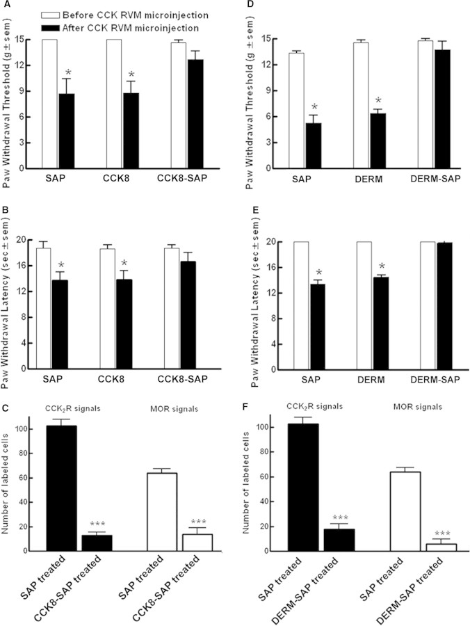
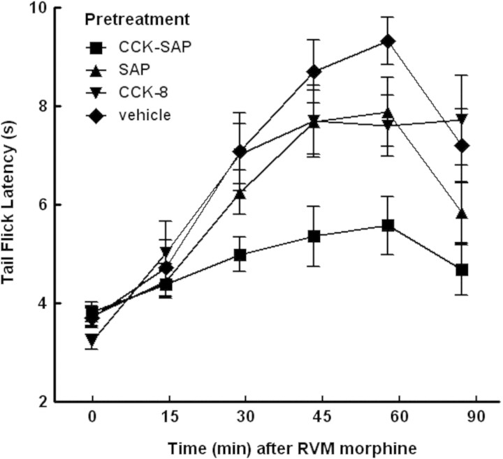
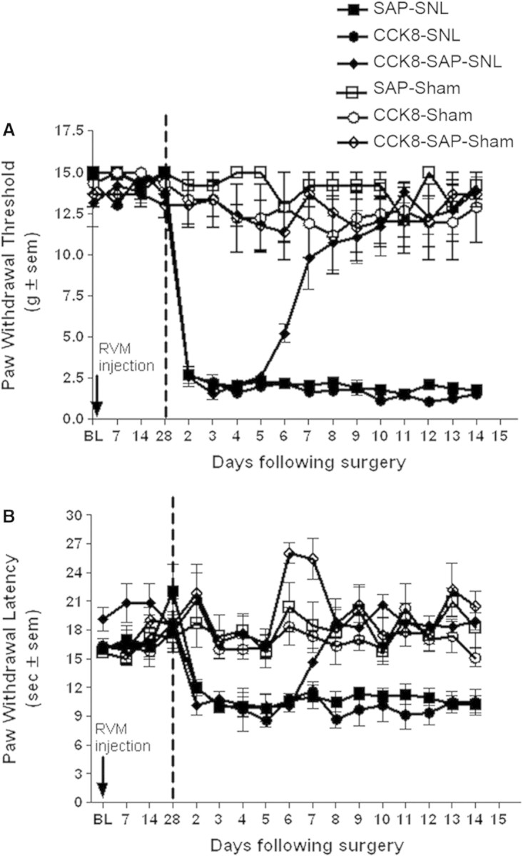
Descending input from the rostral ventromedial medulla (RVM) provides positive and negative modulation of spinal nociceptive transmission and has been proposed to be critical for maintaining neuropathic pain. This study tests the hypothesis that neuropathic pain requires the activity of a subset of RVM neurons that are distinguished by co-expression of mu opioid receptor (MOR) and cholecystokinin type 2 receptor (CCK2). Using male Sprague-Dawley rats, we demonstrate that discrete RVM neurons express MOR and CCK2; over 80% of these cells co-express both receptors. Agonist-directed cell lesion in the RVM with the cytotoxin, saporin, using either CCK-saporin to target CCK receptor expressing cells, or dermorphin-saporin to target MOR expressing cells, resulted in concomitant loss of CCK2 and MOR expressing cells, did not alter the basal sensory thresholds but abolished the hyperalgesia induced by microinjection of CCK into the RVM. The findings suggest that these CCK2-MOR co-expressing RVM neurons facilitate pain and can be directly activated by CCK input to the RVM. Furthermore, lesion of these RVM neurons did not affect the initial development of neuropathic pain in the hind paw upon injury to the sciatic nerve, but the abnormal pain states were short lived such that by about day 9 the sensory thresholds had reverted to pre-injury baselines despite the existing neuropathy. These data support our hypothesis and identify CCK2-MOR co-expressing neurons in the RVM as potential therapeutic targets for neuropathic pain.


Figures

Figure 1
Figure 1
Localization of CCK2 receptor mRNA in the RVM. (A) Representative photomicrograph of neurons containing CCK2 mRNA labeled by digoxigenin-labeled cRNA probe of the rat CCK2 and detected by an anti-digoxigenin antibody and Fast Red stain, counterstained with Mayer's hematoxylin (blue nuclei). Scale bar = 25 μm; (B) Neurons containing CCK2 mRNA detected by a [35S]-labeled cRNA probe for rat CCK2 and counterstained with hematoxylin. High-power micrograph shows two large diameter cells labeled with silver grains surrounding the nuclei; (C) Coronal maps of labeled neurons containing CCK2 mRNA (approximately between −11.3 mm and −10.8 mm from bregma). The tracings are arranged at successive caudal (1) to rostral (8) intervals (120 μm). Each dot denotes one labeled neuron. Red line delineates the approximate location of the RVM including GiA and raphe magnus. Thin black lines delineate the approximate location of facial nuclei (VII), pyramids (py) and LPGi lying between VII and RVM. Gi, nucleus reticularis gigantocellularis; (D) Combined NeuN immunoreactivity/[35S]-labeled cRNA probe for rat CCK2 on the same sections. Silver grains produced by [35S] overlaps with NeuN labelling (brown). NeuN-negative cells are visible as clear somata with green nuclear stain.
Figure 2
Figure 2
Localization of MOR mRNA in the RVM. (A) A representative photomicrograph of neurons containing MOR mRNA in the RVM region using a digoxigenin-labeled cRNA probe of the rat MOR and detected by an anti-digoxigenin antibody and NBT/BCIP (dark blue), counterstained with Mayer's hematoxylin (blue nuclei). Scale bar = 50 μm. (B) Coronal maps of neurons containing MOR mRNA. Tracings are arranged at successive caudal (1) to rostral (8) intervals (120 μm) as for Fig. 1C. Each dot denotes one labeled neuron. Red line delineates the approximate location of the RVM including GiA and raphe magnus. Thin black lines delineate the approximate location of facial nuclei (VII), pyramids (py) and LPGi lying between VII and RVM. Gi, nucleus reticularis gigantocellularis.
Figure 3
Figure 3
Double hybridization histochemistry for CCK2 and MOR transcripts in the RVM. (A) A high magnification image (100× objective) of a pyramidal neuron in the RVM labeled by both [35S]-labeled CCK2 cRNA probe (silver grains) and DIG-labeled MOR cRNA probe (dark blue). Scale bar = 25 μm; (B) A representative map of cells co-labeled for CCK2 and MOR mRNA (closed circles), CCK2 mRNA only (open circles), and MOR mRNA only (crosses) in the RVM. Based on the analysis of nine sections from five rats, over 80% of labeled RVM neurons co-express both MOR and CCK2, about 15% express only CCK2, and very few cells express only MOR. py, pyramid.
Figure 4
Figure 4
Rats received a bilateral microinjection of saporin (SAP), CCK-8(SO3) (CCK8), CCK-8(SO3)-saporin (CCK8-SAP), dermorphin (DERM), or dermorphin-saporin (DERM-SAP) into the RVM. After 28 days, rats were injected with CCK8 (30 ng/0.5 μl, each side) bilaterally into the RVM and were tested for (A and D) paw withdrawal threshold to von Frey probing or (B and E) paw withdrawal latency to radiant heat. (C and F) Effect of RVM pretreatment with SAP or CCK8-SAP (C) and that of RVM pretreatment with SAP or DERM-SAP (F) on the number of CCK2 and MOR expressing neurons in the RVM 28 days after treatment. Positive cells expressing either CCK2 or MOR transcripts were detected by in situ hybridization using digoxigenin-labeled CCK2 or MOR cRNA probe. The cells were mapped and counted using Neurolucida software. Data are expressed as mean ± S.E.M.; *P < 0.05; ***P < 0.001.
Figure 5
Figure 5
Rats (n = 7–12 per group) received bilateral microinjection of water, saporin (SAP), CCK-8(SO3) (CCK8) or CCK-8(SO3)-saporin (CCK8-SAP) into the RVM. After 28 days, rats were injected with morphine (20 μg/1 µl total) bilaterally into the RVM and tested for tail-flick latency at 52°C before and over 90 min after morphine administration. Significant differences in tail flick latencies among treatment groups over time were determined by 2-factor ANOVA. Antinociceptive responses to morphine obtained in rats treated with CCK-SAP in the RVM were significantly [F(5,23) = 7.40; P = 0.0028] lower than those obtained from the vehicle-treated control group.
Figure 6
Figure 6
Male Sprague-Dawley rats received a bilateral microinjection of saporin (SAP) (50 ng/0.5 μl, each side), CCK-8(SO3)(CCK8) (1.5 ng/0.5 μl, each side), or CCK-8(SO3)-saporin (CCK8-SAP)(50 ng/0.5 μl, each side) into the RVM. Paw withdrawal threshold to von Frey probing (A) and paw withdrawal latency to radiant heat (B) were determined prior to RVM injection, and on day 7, 14 and 28 after treatment. After 28 days, the rats were subjected to either L5/L6 SNL or sham surgery. Vertical dashed lines represent time of surgery. Behavioral testing was performed once daily from day 2 to day 14 after SNL or sham surgery. Each group consisted of 4–6 rats.

References

    1. Berg-von der Emde K, Dees WL, Hiney JK, Hill DF, Dissen GA, Costa ME, et al. Neurotrophins and the neuroendocrine brain: different neurotrophins sustain anatomically and functionally segregated subsets of hypothalamic dopaminergic neurons. J Neurosci. 1995;15:4223–37. - PMC - PubMed
    1. Burgess SE, Gardell LR, Ossipov MH, Malan TP Jr, Vanderah TW, Lai J, et al. Time-dependent descending facilitation from the rostral ventromedial medulla maintains, but does not initiate, neuropathic pain. J Neurosci. 2002;22:5129–36. - PMC - PubMed
    1. Chaplan SR, Bach FW, Pogrel JW, Chung JM, Yaksh TL. Quantitative assessment of tactile allodynia in the rat paw. J Neurosci meth. 1994;53:55–63. - PubMed
    1. Fields HL. Nociceptive transmission: the pain system. Science. 1985;228:1522. - PubMed
    1. Fields HL, Heinricher MM, Mason P. Neurotransmitters in nociceptive modulatory circuits. Annu Rev Neurosci. 1991;14:219–45. - PubMed

Publication types

MeSH terms