. 2009 Mar 1;18(5):797-808.
doi: 10.1093/hmg/ddn406. Epub 2008 Dec 2.

Differential requirements for retinal degeneration slow intermolecular disulfide-linked oligomerization in rods versus cones

Affiliations

Differential requirements for retinal degeneration slow intermolecular disulfide-linked oligomerization in rods versus cones

Dibyendu Chakraborty et al. Hum Mol Genet. .

Abstract

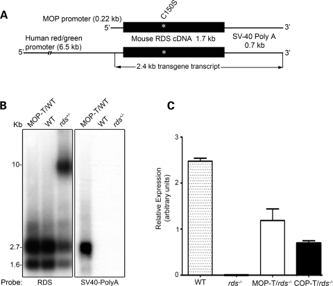
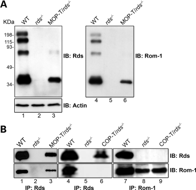
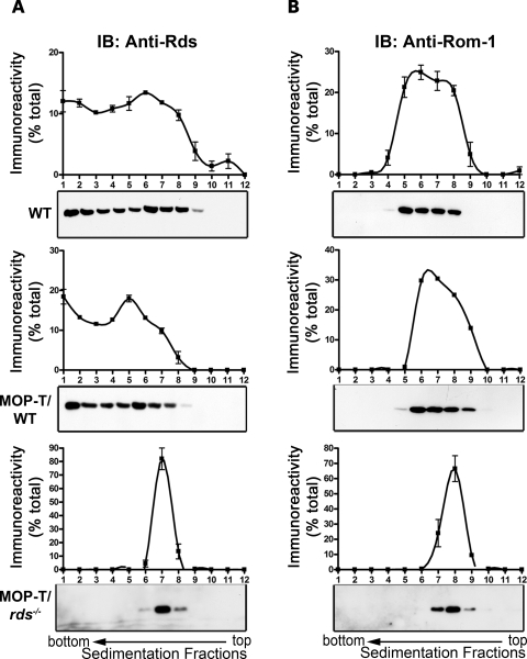
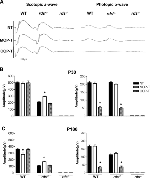
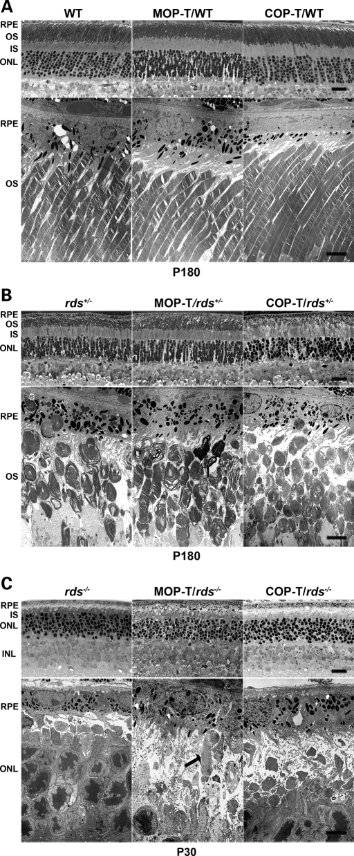
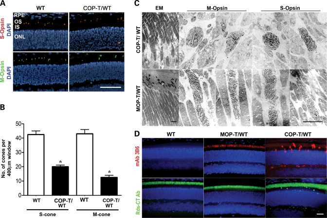
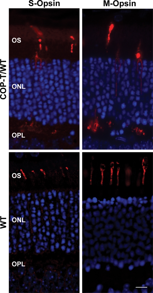
It is commonly assumed that the ultrastructural organization of the rim region of outer segment (OS) discs in rods and lamellae in cones requires functional retinal degeneration slow/rod outer segment membrane protein 1 (Rds/Rom-1) complexes. Cysteine-150 (C150) in Rds has been implicated in intermolecular disulfide bonding essential for functional Rds complexes. Transgenic mice containing the Rds C150S mutation (C150S-Rds) failed to form higher-order Rds oligomers, although interactions between C150S-Rds and Rom-1 occurred in rods, but not in cones. C150S-Rds mice exhibited marked early-onset reductions in cone function and abnormal OS structure. In contrast, C150S-Rds expression in rods partly rescued the rds(+/-) phenotype. Although C150S-Rds was detected in the OSs in rods and cones, a substantial percentage of C150S-Rds and cone opsins were mislocalized to different cellular compartments in cones. The results of this study provide novel insights into the importance of C150 in Rds oligomerization and the differences in Rds requirements in rods versus cones. The apparent OS structural differences between rods and cones may cause cones to be more susceptible to the elimination of higher-order Rds/Rom-1 oligomers (e.g. as mediated by mutation of the Rds C150 residue).


Figures

Figure 1.
Figure 1.
Generation and expression of the C150S-Rds transgene. (A) Schematic diagram of C150S transgene constructs. The transgene is composed of the full-length mouse RDS cDNA with the C150S mutation followed by an SV40 poly-A tail and directed either to rods by the MOP or to cones by the human red/green opsin promoter (COP). (B) Northern blot analysis of whole retinal RNA extracts (10 µg/sample) taken from 1-month-old MOP-T transgenic, non-transgenic WT littermates and rds+/− mice. Two different probes were hybridized to the membrane: one from exon 1 of the RDS cDNA that recognizes both endogenous and transgene transcripts (left) and an SV40 poly-A probe that recognizes the transgenic message only (right). (C) qRT–PCR was used to measure levels of total RDS message from WT, rds−/−, MOP-T/rds−/− and COP-T/rds−/− retinas. Analysis was performed on retinas from 3- to 4-week-old mice using primer specific to the endogenous RDS gene.
Figure 2.
Figure 2.
C150S-Rds protein is stably formed in rods and cones. (A) Retinal extracts were isolated from 1-month-old MOP-T animals in the rds−/− background and controls and were analyzed by non-reducing western blot. The blot was sequentially probed with anti-Rds-CT (left), anti-actin (left bottom) and anti-Rom-1-CT antibodies (right). (B) Western blot analysis of anti-Rds-CT (left and middle) or anti-Rom-1-CT (right) immunoprecipitants from retinal extracts of WT, rds−/−, MOP-T/rds−/− and COP-T/rds−/− mice. Blots were probed with anti-Rds-CT (top) and anti-Rom-1-CT antibodies (bottom). Both MOP-T and COP-T Rds are expressed, although Rom-1 binds to MOP-T, but not to COP-T, Rds.
Figure 3.
Figure 3.
The effect of C150S-Rds on the pattern of complex assembly in rods. Non-reducing velocity sedimentation was performed on WT, MOP-T/WT and MOP-T/rds−/− retinal extracts in the presence of NEM and Triton X-100. Fractionated gradients (–12) were collected and evaluated by western blot using anti-Rds-CT (A) and anti-Rom-1-CT (B) antibodies. Image analysis was performed on blots from three independent experiments for each genotype and corresponding densitometry plots (mean±S.E.M.) are presented to show the distribution patterns of Rds and Rom-1 complexes. The distribution of these complexes is similar in MOP-T/WT and WT retinal extracts. However, in the absence of native Rds, C150S Rds was unable to make higher-order or intermediate complexes but retained the ability to make tetramers (bottom).
Figure 4.
Figure 4.
Functional defects associated with the C150S mutation. (A) Scotopic (left) and photopic (right) ERG wave forms from 1-month-old MOP-T, COP-T and non-transgenic animals in the WT, rds+/− and rds−/− backgrounds. (B) Quantitation of scotopic (left) and photopic (right) ERG amplitudes of 1-month-old C150S mice and controls in the WT, rds+/− and rds−/− backgrounds. At P30 a significant cone defect is associated with the expression of C150S-Rds in WT and rds+/− backgrounds and no rod or cone function was seen when C150S is expressed in the rds−/− background. (C) ERG analysis of 6-month-old C150S mice in all genetic backgrounds. Expression of C150S (MOP-T) exerted a late-onset functional defect in rods. However, expression of C150S-Rds in cones (COP-T) continued to dramatically affect cone function. Shown are means±S.E.M. from 5–7 mice. Significant differences as measured by one-way ANOVA (P < 0.001) between transgenic and non-transgenic littermates are marked with asterisks (*).
Figure 5.
Figure 5.
C150S improves rod photoreceptor OS structure in rds+/− retina. Shown are representative light (top) and electron (bottom) microscopy from retinal sections of MOP-T, COP-T and non-transgenic controls in the WT (A), rds+/− (B) and rds−/− (C) backgrounds. Eyes presented in A and B were taken from mice at P180 and in C from mice at P30. Expression of C150S-Rds had no effect on rod OS structure in the WT background and led to partial improvement in OS structure in the rds+/− background. As expected, no change in rod OS structure was noticed in COP-T/rds+/− or COP-T/WT retinas compared with non-transgenic controls. (C) Except in rare instances (arrow) C150S-Rds was not sufficient to support OS formation in the absence of endogenous Rds. RPE, retinal pigment epithelium; OS, outer segment; IS, inner segment; ONL, outer nuclear layer; INL, inner nuclear layer. Scale bar, 4 µm (EM) and 20 µm (light).
Figure 6.
Figure 6.
(A) Paraffin-embedded retinal sections from WT and COP-T/WT mice were labeled with anti-S-opsin (red) or anti-M-opsin (green) and counterstained with DAPI (blue). RPE, retinal pigment epithelium; OS, outer segment; IS, inner segment; ONL, outer nuclear layer. Scale bar, 50 µm. (B) The number of cones in retinas of COP-T/WT mice at P30 was significantly decreased compared with non-transgenic controls (mean±S.E.M. n = 3, *=P < 0.001 by Student’s t-test). (C) Immunogold labeling of M- and S-cones (with the corresponding opsin antibodies) in COP-T/WT (top) and MOP-T/WT (bottom) retinas at P30. Expression of C150S Rds in cones (COP-T/WT) results in shorter, abnormal cone lamellae compared with the normal cones seen in the MOP-T/WT. Scale bars: EM, 4 µm; IM, 2 µm. (D) Paraffin-embedded retinal sections were labeled with mAb 3B6 to visualize C150S Rds or with anti-Rds-CT polyclonal antibody to visualize both endogenous and transgenic Rds. C150S-Rds is properly localized to the OS of MOP-T/WT retinas, whereas mislocalized to the inner segments, outer nuclear layer and the outer plexiform layer of COP-T/WT retinas. Scale bar, 20 µm.
Figure 7.
Figure 7.
Paraffin-embedded sections from COP-T mice and non-transgenic littermates were collected at P30 and stained with anti-S-opsin or anti-M-opsin to label the cone opsin proteins (left and right). In the presence of the C150S-Rds protein in the COP-T retina (top), both S- and M-opsin are distributed throughout the photoreceptor in contrast to the WT (bottom) in which the protein is restricted to the OS. Scale bar 10 µm.

References

    1. Corless J.M., Fetter R.D., Costello M.J. Structural features of the terminal loop region of frog retinal rod outer segment disk membranes. I. Organization of lipid components. J. Comp. Neurol. 1987;257:1–8. - PubMed
    1. Eckmiller M.S. Cone outer segment morphogenesis: taper change and distal invaginations. J. Cell Biol. 1987;105:2267–2277. - PMC - PubMed
    1. Roof D.J., Heuser J.E. Surfaces of rod photoreceptor disk membranes: integral membrane components. J. Cell Biol. 1982;95:487–500. - PMC - PubMed
    1. Chuang J.Z., Zhao Y., Sung C.H. SARA-regulated vesicular targeting underlies formation of the light-sensing organelle in mammalian rods. Cell. 2007;130:535–547. - PMC - PubMed
    1. Arikawa K., Molday L.L., Molday R.S., Williams D.S. Localization of peripherin/Rds in the disk membranes of cone and rod photoreceptors: relationship to disk membrane morphogenesis and retinal degeneration. J. Cell Biol. 1992;116:659–667. - PMC - PubMed

Publication types

MeSH terms