. 2008 Dec;20(12):3300-11.
doi: 10.1105/tpc.108.062919. Epub 2008 Dec 2.

Truncation of a protein disulfide isomerase, PDIL2-1, delays embryo sac maturation and disrupts pollen tube guidance in Arabidopsis thaliana

Affiliations

Truncation of a protein disulfide isomerase, PDIL2-1, delays embryo sac maturation and disrupts pollen tube guidance in Arabidopsis thaliana

Huanzhong Wang et al. Plant Cell. 2008 Dec.

Abstract

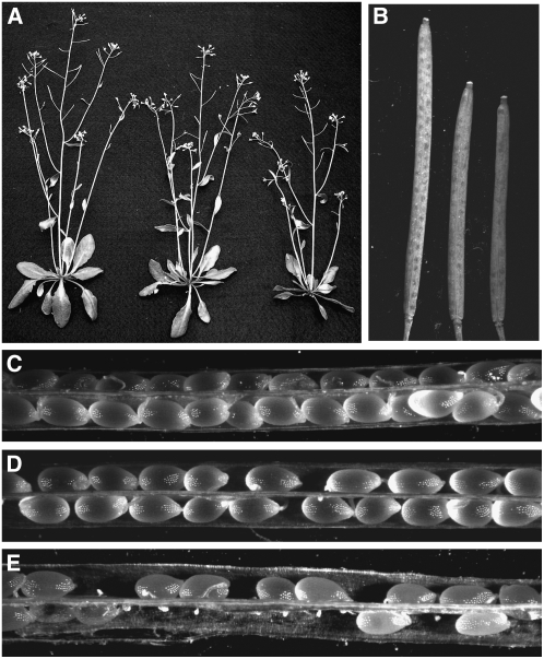
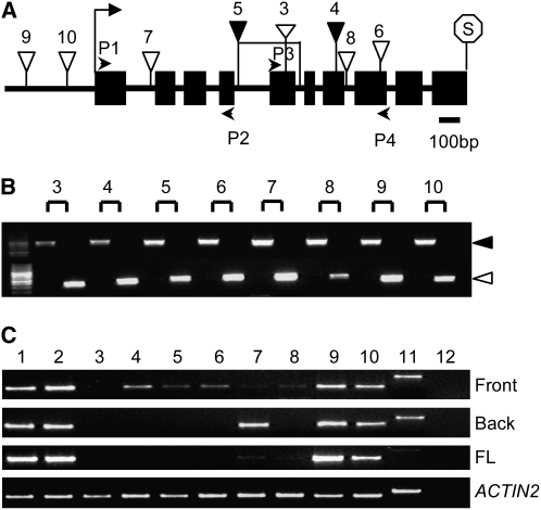
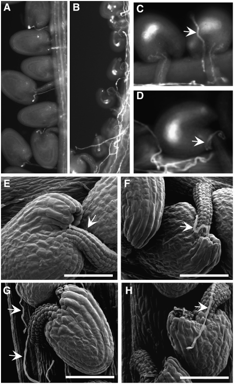
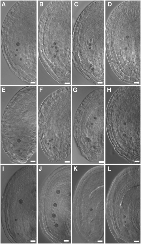
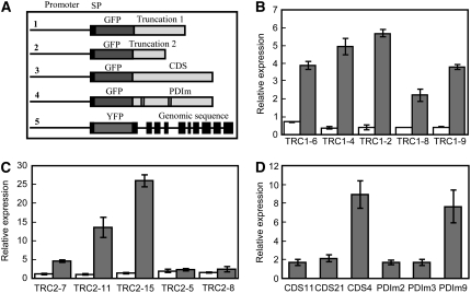
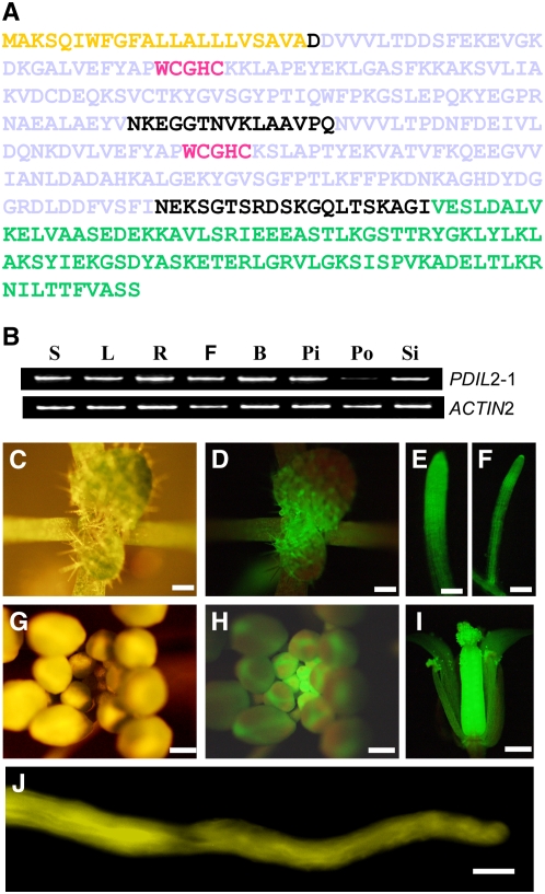
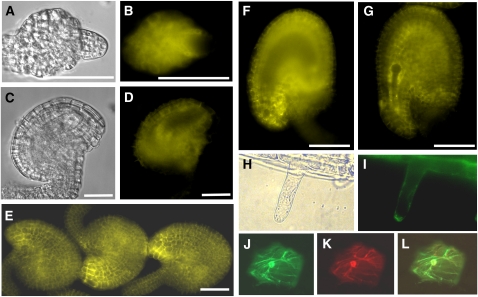
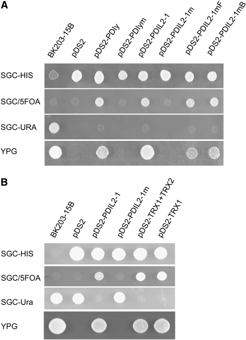
Pollen tubes must navigate through different female tissues to deliver sperm to the embryo sac for fertilization. Protein disulfide isomerases play important roles in the maturation of secreted or plasma membrane proteins. Here, we show that certain T-DNA insertions in Arabidopsis thaliana PDIL2-1, a protein disulfide isomerase (PDI), have reduced seed set, due to delays in embryo sac maturation. Reciprocal crosses indicate that these mutations acted sporophytically, and aniline blue staining and scanning electron microscopy showed that funicular and micropylar pollen tube guidance were disrupted. A PDIL2-1-yellow fluorescent protein fusion was mainly localized in the endoplasmic reticulum and was expressed in all tissues examined. In ovules, expression in integument tissues was much higher in the micropylar region in later developmental stages, but there was no expression in embryo sacs. We show that reduced seed set occurred when another copy of full-length PDIL2-1 or when enzymatically active truncated versions were expressed, but not when an enzymatically inactive version was expressed, indicating that these T-DNA insertion lines are gain-of-function mutants. Our results suggest that these truncated versions of PDIL2-1 function in sporophytic tissues to affect ovule structure and impede embryo sac development, thereby disrupting pollen tube guidance.


Figures

Figure 1.
Figure 1.
Vegetative and Reproductive Development of Wild-Type and Line 5 Plants. (A) Thirty-five-day-old homozygous plants (right) and heterozygous plants (middle) show a slight reduction in growth relative to wild-type plants (left). (B) Mature siliques from the homozygous (right) and heterozygous mutant (middle) are shorter than wild-type siliques (left). (C) Dissected mature wild-type siliques with full seed set. (D) Siliques from the heterozygous plants with reduced seed set. (E) Siliques from homozygous plants have severely reduced seed set and obvious undeveloped ovules. Similar results were seen with line 4.
Figure 2.
Figure 2.
Gene Structure and T-DNA Insertion Analyses of PDIL2-1. (A) PDIL2-1 gene structure and T-DNA insertion sites. P1, P2, P3, and P4 indicate position of primers used to check expression. Start and stop codons are noted with an arrow and octagon, respectively. Black boxes and black lines represent exons and introns, respectively. (B) PCR identification of homozygotes in the indicated T-DNA insertion lines. For each line, the left lane is a wild-type plant having only a gene-specific band (black arrow), and the right lane is an insertion line, having only a T-DNA–specific band (white arrow). (C) RT-PCR analyses. PDIL2-1 expression was examined using primers covering different parts of the gene (primers P1 and P2, front; or primers P3 and P4, back) or full-length (FL) cDNA. ACTIN2 expression was used as a control. The numbers shown in (A) to (C) represent (1) Ws4 wild type, (2) Col-0 wild type, (3) FLAG_015D01, (4) FLAG_427G04, (5) SALK_046705, (6) SALK_082179, (7) SALK_135268c, (8) SALK_148421, (9) SALK_097094, (10) CS859015, (11) gDNA control, and (12) water control.
Figure 3.
Figure 3.
Line 5 Homozygous Plants Show Disrupted Pollen Tube Guidance. (A) to (D) Decolorized aniline blue staining of 3 d after pollination (DAP) pistils. (A) Wild-type siliques showing fertilized ovules and pollen tube growth. (B) Mutant siliques showing randomly growing pollen tubes and undeveloped ovules. (C) A representative mutant ovule with pollen tube (arrow) growing on the ovule surface. (D) An ovule with pollen tube (arrow) growing around the funiculus without targeting the micropyle. (E) and (F) Scanning electron micrographs of 2-DAP pistils. Two ovules from a wild-type plant show normal pollen tube (arrow) growth and correct targeting to the micropyle. (G) A mutant ovule with a pollen tube (arrow) not targeted to the funiculus or micropyle. (H) A mutant ovule with a pollen tube (arrow) growing on the surface. Bars = 50 μm in (E) to (H).
Figure 4.
Figure 4.
Embryo Sac Development in Mutant Line 5 and PDIL2-1 Transgenic Plants. Heterozygous ([A] to [D]) and homozygous ([E] to [H]) ovules of line 5. Normal embryo sac ([A] and [E]), embryo sac with eight nuclei ([B] and [F]), no cellularization, four nuclei ([C] and [G]), and two nuclei ([D] and [H]). (I) to (L) Representative embryo sac of CDS, truncation 1, and truncation 2 transgenic lines (see Figure 5A), with normal embryo sac (I), four nuclei (J), one nuclei (K), and two nuclei (L), respectively. Bars = 20 μm.
Figure 5.
Figure 5.
Construct Structures and Real-Time Quantitative PCR Analyses of Transgenic Lines. (A) Schematic structure of five transformation constructs. SP denotes signal peptide. Truncation 1 and Truncation 2 (TRC1 and TRC2) correspond to insertion sites of line 4 and line 5. CDS denotes the coding sequence of PDIL2-1. PDIm denotes the coding sequence of PDIL2-1 with both active sites mutated. (B) to (D) Real-time quantitative PCR analyses of the transgenic lines. Endogenous and total PDIL2-1 expression (endogenous and from the transgene) were examined in truncation 1 (B), truncation 2 (C), or CDS and PDIm (D) transgenic plants. White bars in (B) and (C) represent endogenous expression, and gray bars represent total expression. Error bars represent ±sd of four technical replicates.
Figure 6.
Figure 6.
Protein Structure and Expression Pattern of PDIL2-1. (A) Protein sequence of PDIL2-1. Yellow denotes the signal peptide (SP), gray denotes the two thioredoxin domains (TRX), red denotes the active sites, and green denotes the D domain. (B) Expression of PDIL2-1 in different organs was examined by RT-PCR using gene-specific and control (ACT2) primers. S, seedlings; L, leaves; R, roots; F, opened flowers; B, vlosed flower buds; Pi, unfertilized pistils; Po, pollen; Si, siliques. (C) to (J) Expression pattern determined by YFP fusion reporter (ProPDIL2-1:SP-eYFP-PDIL2-1). (C) and (G) Vegetative and reproductive apical region. (D) and (H) The same region under UV light (GFP filter). (E), (F), and (I) Expression of PDIL2-1 in lateral and main roots and in a flower, respectively. (J) In vitro–germinated pollen tube. Bars = 500 μm in (C), (D), and (G) to (I), 100 μm in (E) and (F), and 10 μm in (J).
Figure 7.
Figure 7.
PDIL2-1 Expression in Ovules and Subcellular Localization. (A) and (B) Ovule of early stage 12 flowers under differential interference contrast (DIC) (A) or YFP channel (B). YFP signal is visible in integument cells and nucellus cells. (C) and (D) Ovule of late stage 12 flowers under DIC (C) or YFP channel (D). YFP is visible in integument cells. (E) Mature ovules of stage 13 flowers. YFP signal is visible in integument cells and is highly accumulated at the micropyle. (F) An ovule of stage 13 flowers. Focus is on the plane of the embryo sac to show no signal inside of the embryo sac. (G) A fertilized ovule with no YFP signal in the developing embryo. (H) Root hair under transmitted light. (I) ProPDIL2-1:SP-YFP-PDIL2-1 expression in root hairs. (J) GFP-KDEL, an ER marker. (K) PDIL2-1-tdTomato red fusion protein. (L) Overlay of (J) and (K). Bars = 20 μm in (A) to (D) and 50 μm in (E) to (G).
Figure 8.
Figure 8.
PDIL2-1 Has PDI Activity. (A) Yeast complementation analysis showing that PDIL2-1 is a functional isomerase. (B) Truncated PDIL2-1 also showed enzyme activity in yeast. In both panels, the left column lists the medium used for growth. The first column is yeast strain BK203-15B, and the second column is empty pDS2 vector. Transformants with different constructs are marked at the top. The wild-type yeast PDIy and mutated PDIym (with both CGHC boxes mutated to SGHS) were used as positive and negative controls, respectively. PDIL2-1 and PDIL2-1m were constructed similarly to the yeast PDI constructs. PDIL2-1mF and PDIL2-1mB are different mutations of PDIL2-1, with either the first or second CGHC boxes mutated. TRX1 and TRX2 represent the two TRX domains. Note that only constructs with a functional PDI can grow on YPG plates.

References

    1. Alandete-Saez, M., Ron, M., and McCormick, S. (2008). GEX3, expressed in the male gametophyte and in the egg cell of Arabidopsis thaliana, is essential for micropylar pollen tube guidance and plays a role during early embryogenesis. Molecular Plant 1 586–598. - PubMed
    1. Alonso, J.M., et al. (2003). Genome-wide insertional mutagenesis of Arabidopsis thaliana. Science 301 653–657. - PubMed
    1. Baker, S.C., Robinson-Beers, K., Villanueva, J.M., Gaiser, J.C., and Gasser, C.S. (1997). Interactions among genes regulating ovule development in Arabidopsis thaliana. Genetics 145 1109–1124. - PMC - PubMed
    1. Bhatt, A.M., Lister, C., Page, T., Fransz, P., Findlay, K., Jones, G.H., Dickinson, H.G., and Dean, C. (1999). The DIF1 gene of Arabidopsis is required for meiotic chromosome segregation and belongs to the REC8/RAD21 cohesin gene family. Plant J. 19 463–472. - PubMed
    1. Boavida, L.C., and McCormick, S. (2007). Temperature as a determinant factor for increased and reproducible in vitro pollen germination in Arabidopsis thaliana. Plant J. 52 570–582. - PubMed

Publication types

MeSH terms

Associated data