Comparative Study
. 2008 Dec 15;181(12):8576-84.
doi: 10.4049/jimmunol.181.12.8576.

IL-12 produced by dendritic cells augments CD8+ T cell activation through the production of the chemokines CCL1 and CCL17

Affiliations
Comparative Study

IL-12 produced by dendritic cells augments CD8+ T cell activation through the production of the chemokines CCL1 and CCL17

Curtis J Henry et al. J Immunol. .

Abstract

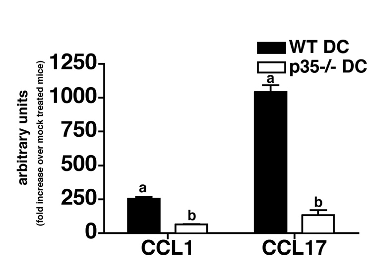
IL-12 family members are an important link between innate and adaptive immunity. IL-12 drives Th1 responses by augmenting IFN-gamma production, which is key for clearance of intracellular pathogens. IL-23 promotes the development of IL-17-producing CD4(+) T cells that participate in the control of extracellular pathogens and the induction of autoimmunity. However, recent studies have shown that these cytokines can modulate lymphocyte migration and cellular interactions. Therefore, we sought to determine the individual roles of IL-12 and IL-23 in naive CD8(+) T cell activation by addressing their ability to influence IFN-gamma production and cellular interaction dynamics during priming by Listeria monocytogenes-infected dendritic cells (DC). We found that IL-12 was the major cytokine influencing the level of IFN-gamma production by CD8(+) T cells while IL-23 had little effect on this response. In addition, we observed that IL-12 promoted longer duration conjugation events between CD8(+) T cells and DC. This enhanced cognate interaction time correlated with increased production of the chemokines CCL1 and CCL17 by WT but not IL-12-deficient DC. Neutralization of both chemokines resulted in reduced interaction time and IFN-gamma production, demonstrating their importance in priming naive CD8(+) T cells. Our study demonstrates a novel mechanism through which IL-12 augments naive CD8(+) T cell activation by facilitating chemokine production, thus promoting more stable cognate interactions during priming.


Figures

FIGURE 1
FIGURE 1. IL-12, but not IL-23 augments IFN-γ production by naïve CD8+ T cells
A, CFSE-stained OT-1 T cells were primed by Listeria-infected OVA-pulsed wild-type, p35−/−, or p40−/− DC and IFN-γ production was determined by ICS staining on day 3 of priming. IFN-γ MFI of T cells is denoted in the upper left corner. Contour plots are representative of 10 independent experiments. B, The combined data from eight independent flow cytometric experiments is graphed. The solid line indicates the amount of IFN-γ T cells produced when primed in the absence of OVA peptide. The mean ± the SD is shown. C, IFN-γ production by CD8+ T cells during priming measured by ELISA. CD8+ T cells were primed by either wild-type, p35−/−, or p40−/− DC and the amount of IFN-γ that accumulated over three days was determined via ELISAs. The average concentration of IFN-γ from three independent experiments is graphed. D, CFSE-stained OT-1 T cells were primed by Listeria-infected OVA-pulsed DC in the presence of neutralizing antibodies against IL-23 (anti-p19) or IL-12/23 (anti-p40) and IFN-γ production was determined by ICS staining on day 3 of priming. Contour plots are representative of eight independent experiments. E, The combined data from 10 independent experiments is graphed. The solid line indicates the amount of IFN-γ T cells produced when primed by wild-type DC in the absence of OVA peptide. The mean ± the SD is shown. Significance compared to the Rat Ig or WT DC controls was determined using a Student’s t-test with *, p < 0.05; **, p < 0.01; ***, p < 0.001.
FIGURE 2
FIGURE 2. IL-12 promotes long duration interactions with DC
A, OT-1 were primed by Listeria-infected, OVA-pulsed DC for 48 hours in vitro with control or anti-p40 neutralizing antibodies. The duration of interaction between T cells and DC were determined via time lapse video microscopy. B, OT-1 were primed by wild-type, p35−/−, or p40−/− DC as described above and the duration of interaction between T cells and DC were determined via time lapse video microscopy. Dissociation curves are combined data from at least three independent experiments per condition. The solid line in A and B represents 50% of the interactions analyzed. Significance compared to the WT DC + OVA control samples was determined by Cox Proportional Hazard Regression Analysis. The neutralization of IL-12/23 (anti-p40) resulted in a significant decrease in the duration of interaction of OT-1 (p < 0.01) with DC, which was further reduced in the absence of IL-12 (p35−/− DC; p < 0.001) or IL-12/23 (p40−/− DC; p < 0.001).
Figure 3
Figure 3. IL-12 increases the expression of CCL1 and CCL17 message and secretion in Lm-infected DC
Wild-type, p35−/−, p40−/−, and IL-12R−/− DC were mock treated or infected with Lm (MOI 1), and RNA was isolated from these DC at 24 hours post infection. A, CCL1 and CCL17 message levels were determined via real-time PCR. Data represents combined results from 3 independent experiments and the mean ± SD is shown. (a) indicates that samples was significantly different (b). B and C, Wild-type, p35−/−, and IL-12R−/− DC were infected with Lm at an MOI of 1. Chemokine secretion was measured 24h later by ELISA. In some experiments recombinant IL-12 was added back to IL-12 deficient DC (p35−/−) in order to determine the effects this treatment had on CCL17 (B) and CCL1 production (C). Significance was determined compared to the WT DC control using a Student’s t-test with *, p < 0.05; **, p < 0.01; ***, p < 0.001.
FIGURE 4
FIGURE 4. CCL1 and CCL17 augment T cell/DC interaction time and IFN-γ production by CD8+ T cells
A and B OT-1 and P14 were primed by OVA or GP33-41 pulsed Listeria-infected DC for 72 hours in vitro with control or anti-CCL1/CCL17 neutralizing antibodies. The duration of interaction between T cells and DC were determined via time lapse video microscopy. Dissociation curves are combined data from at least three independent experiments per condition. The solid line represents 50% of the interactions analyzed. Significance compared to the control samples was determined by Cox Proportional Hazard Regression Analysis. The neutralization of CCL1/17 resulted in a significant decrease in the duration of interaction of OT-1 (p<0.01) and P14 (p<0.01) with DC compared to controls. C, OT-1 and P14 were primed by Listeria-infected, OVA or GP33-41 peptide pulsed DC for 72 hours in vitro. Control, anti-CCL1, anti-CCL17, or a combination of anti-CCL1/17 neutralizing antibodies was added when T cells were added with DC. D, The IFN-γ MFI of T cells was determined at 72 hours via ICS staining. Compiled data represent 8 independent experiments for the OT-1 T cells and 4 independent experiments for the P14 T cells. The relative statistical significance of either OT-1 or P14 CD8+ T cell responses were determined compared to the same T cell in the absence of neutralization using a Student’s t-test with *, p < 0.05; **, p < 0.01; ***, p < 0.001.
Figure 5
Figure 5. CCL1 and CCL17 do not augment IFN-γ production by CD4+ T cells
OT-1 and OT-2 T cells were primed by Lm-infected DC in the presence or absence of anti-p40, anti-CCL1, anti-CCL17, or a combination of anti-CCL1/17 neutralizing antibodies in vitro for 3 days. On day 3 of priming, supernatants were filtered and assayed for the concentration of IFN-γ via ELISAs. A, Histograms represent combined data from 5 independent experiments for OT-1 and B, three independent experiments for OT-2 T cells. Significance of neutralized samples was calculated in comparison to control OT-1 CD8+ T cells or OT-2 CD4+ T cells without neutralization using a Student’s t-test with *, p < 0.05; **, p < 0.01; ***, p < 0.001.
Figure 6
Figure 6. IL-12 augments CCL1 and CCL17 message in DC during Lm infection in vivo
Wild-type and IL-12 deficient (p35−/−) DC were mock treated (PBS) or infected with 1 LD50 of Lm intravenously. At 18 hours post infection, RNA was isolated from DC enriched from the lymph nodes of treated mice. Four mice per group were analyzed and data represent the mean ± SD. CCL1 and CCL17 message levels were determined via real-time PCR. Significance was assessed compared to DC isolated from WT mice using a Student’s t-test for each gene with *, p < 0.05; **, p < 0.01; ***, p < 0.001.

References

    1. Kang SS, Allen PM. Priming in the presence of IL-10 results in direct enhancement of CD8+ T cell primary responses and inhibition of secondary responses. J. Immunol. 2005;174:5382–5389. - PubMed
    1. Sallusto F, Lanzavecchia A. The instructive role of dendritic cells on T-cell responses. Arthritis Res. 4 Suppl. 2002;3:S127–S132. - PMC - PubMed
    1. Chang J, Cho JH, Lee SW, Choi SY, Ha SJ, Sung YC. IL-12 priming during in vitro antigenic stimulation changes properties of CD8 T cells and increases generation of effector and memory cells. J. Immunol. 2004;172:2818–2826. - PubMed
    1. Chang J, Choi SY, Jin HT, Sung YC, Braciale TJ. Improved effector activity and memory CD8 T cell development by IL-2 expression during experimental respiratory syncytial virus infection. J. Immunol. 2004;172:503–508. - PubMed
    1. Langenkamp A, Messi M, Lanzavecchia A, Sallusto F. Kinetics of dendritic cell activation: impact on priming of TH1, TH2 and nonpolarized T cells. Nat. Immunol. 2000;1:311–316. - PubMed

Publication types

MeSH terms