. 2009 May 7;113(19):4667-76.
doi: 10.1182/blood-2008-07-171637. Epub 2008 Dec 2.

Targeted inhibition of the immunoproteasome is a potent strategy against models of multiple myeloma that overcomes resistance to conventional drugs and nonspecific proteasome inhibitors

Affiliations

Targeted inhibition of the immunoproteasome is a potent strategy against models of multiple myeloma that overcomes resistance to conventional drugs and nonspecific proteasome inhibitors

Deborah J Kuhn et al. Blood. .

Abstract

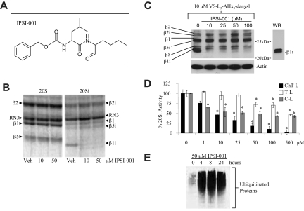
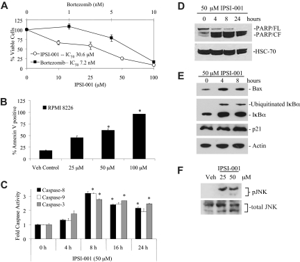
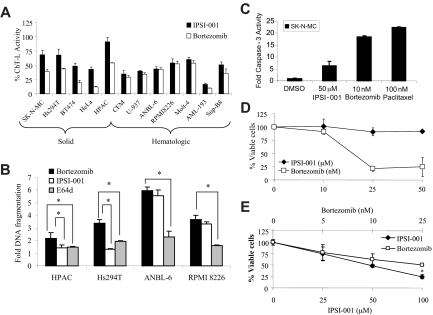
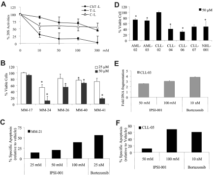
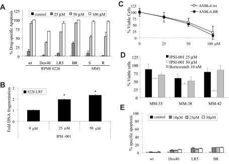
Proteasome inhibition is a validated strategy for therapy of multiple myeloma, but this disease remains challenging as relapses are common, and often associated with increasing chemoresistance. Moreover, nonspecific proteasome inhibitors such as bortezomib can induce peripheral neuropathy and other toxicities that may compromise the ability to deliver therapy at full doses, thereby decreasing efficacy. One novel approach may be to target the immunoproteasome, a proteasomal variant found predominantly in cells of hematopoietic origin that differs from the constitutive proteasome found in most other cell types. Using purified preparations of constitutive and immunoproteasomes, we screened a rationally designed series of peptidyl-aldehydes and identified several with relative specificity for the immunoproteasome. The most potent immunoproteasome-specific inhibitor, IPSI-001, preferentially targeted the beta1(i) subunit of the immunoproteasome in vitro and in cellulo in a dose-dependent manner. This agent induced accumulation of ubiquitin-protein conjugates, proapoptotic proteins, and activated caspase-mediated apoptosis. IPSI-001 potently inhibited proliferation in myeloma patient samples and other hematologic malignancies. Importantly, IPSI-001 was able to overcome conventional and novel drug resistance, including resistance to bortezomib. These findings provide a rationale for the translation of IPSIs to the clinic, where they may provide antimyeloma activity with greater specificity and less toxicity than current inhibitors.


Figures

Figure 1
Figure 1
IPSI-001 inhibits the immunoproteasome. (A) Chemical structure of IPSI-001. (B) Competitive binding experiment to determine subunit binding profile of IPSI-001 in purified proteasomes from bovine pituitaries (20S) and spleen (20Si). Purified proteasomes were incubated with 10 or 50 μM IPSI-001 or vehicle (Veh) for 20 minutes, and then [14C]-3,4-dichloroisocoumarin was added for an additional 30 minutes. Proteasomes were then separated by denaturing gel electrophoresis and the binding profile was identified using autoradiography. Subunit identifications were made based on the known electrophoretic migration patterns of immunoproteasome and constitutive proteasome subunits. (C) Competitive binding experiment to determine the subunit binding profile of IPSI-001 in ANBL-6 cells. Cells were treated for 4 hours with IPSI-001, followed by addition of the VS-L3-AHx3-dansyl reagent that binds to all unoccupied catalytic subunits. Protein extracts were then separated by denaturing gel electrophoresis and probed by Western blotting using an antidansyl antibody to visualize binding patterns. Antiactin was used as a loading control and anti-β1i was used to confirm the relative position of β1i. (D) In vitro assay for the 3 major catalytic activities of the proteasome in ANBL-6 cell lysates (5 μg) exposed to increasing concentrations of IPSI-001 in triplicate. Representative data are shown as the mean plus or minus standard deviation (SD) from 3 independent experiments. *P < .05 for proteasome inhibitory activity over control. (E) Western blot analysis of the accumulation of polyubiquitinated proteasome substrates in response to 50 μM IPSI-001 treatment in ANBL-6 cells over a period of 24 hours.
Figure 2
Figure 2
Cell death and molecular changes associated with IPSI-001. (A) WST-1 cytotoxicity assay in ANBL-6 cells treated for 18 hours with bortezomib or IPSI-001. The data shown are the mean plus or minus SD from 3 independent experiments. (B) Flow cytometric analysis was performed after annexin V staining in RPMI 8226 cells treated for 24 hours with the indicated concentrations of IPSI-001, with each experiment performed in duplicate. Error bars indicate SD. *P < .05 comparing treated cells over vehicle control. (C) Representative results from 2 independent experiments of a colorimetric activity assay for caspases in RPMI 8226 myeloma cell lysate (100 μg) treated for up to 24 hours with 50 μM IPSI-001. The data shown are the mean plus or minus SD from duplicate experiments. *P < .05 for fold caspase activity over baseline. (D) Western blot analysis for the late-stage apoptotic marker PARP, where the cleaved fragment (CF) represents apoptotic cells compared with the full-length (FL) PARP, which is present in healthy, nonapoptotic ANBL-6 cells. HSC-70 levels were used as a loading control. (E) Protein expression of Bax and IκBα in RPMI 8226 cells after incubation with 50 μM IPSI-001 for 24 hours. Actin levels were used as a loading control. (F) Protein expression of phosphorylated JNK (phospho-JNK) and total JNK in RPMI 8226 cells treated with 25 or 50 μM IPSI-001 for 24 hours.
Figure 3
Figure 3
IPSI-001 preferentially inhibits the proteasome and induces death in lymphoid-derived cells. (A) The ChT-L activity was measured in vitro in solid tumor (SK-N-MC, Hs294T, BT474, HeLa, and HPAC; lanes 1-5) and hematologic cancer cells (CCRF-CEM, U-937, ANBL-6, RPMI 8226, MOLT-4, AML193, and SupB8; lanes 6-12) by exposing lysates to 50 μM IPSI-001 or 10 nM bortezomib for 15 minutes, followed by incubation with the specific ChT-L activity substrate. The data shown are the mean plus or minus SD from experiments performed in triplicate. (B) HPAC pancreatic adenocarcinoma cells, Hs294T melanoma cells, and ANBL-6 and RPMI 8226 human myeloma cells were exposed to vehicle, 10 nM bortezomib, 25 μM E64d, or 50 μM IPSI-001 for 24 hours. Apoptosis was evaluated using an enzyme-linked immunosorbent assay that detects apoptotic nuclear DNA fragmentation. Cell death was expressed as the fold increase in apoptosis over the vehicle control, which was arbitrarily set at 1.0. Representative data are shown from 3 independent experiments. *P < .05 comparing the inhibitors used. (C) Human SK-N-MC cells were treated for 24 hours with the indicated agents and then cell lysates (30 μg) were measured for caspase-3 activation using a fluorogenic substrate. Representative data are shown from 2 independent experiments, each performed in triplicate, as the mean plus or minus SD. (D) Human umbilical vein endothelial cells (HUVECs) were plated until they were 70% to 80% confluent, and then treated with increasing concentrations of IPSI (μM) or bortezomib (nM) for 24 hours, followed by evaluation by WST-1 for cell viability in triplicate. The data shown are the mean plus or minus SD from experiments performed in triplicate. (E) Human peripheral blood mononuclear cells (PBMCs) were plated and exposed to IPSI-001 and bortezomib. Viable cells were assayed 24 hours later using WST-1 reagent. *P < .05 for IPSI-001 treatment compared with bortezomib.
Figure 4
Figure 4
Multiple myeloma and other primary samples from patients with hematologic malignancies are sensitive to IPSI-001. (A) Cellular extracts (5 μg) from CD33+ acute myeloma leukemia (AML-01) patient cells were exposed to increasing doses of IPSI-001 and proteasome inhibition was assessed using fluorogenic substrates for the ChT-L, T-L, and C-L activities of the proteasome in triplicate. These data are similar to experiments performed in 3 additional CD19+ patient samples. The data shown are the mean plus or minus SD. (B) Purified CD138+ plasma cells from patient samples were treated with 25 and 50 μM IPSI-001 for 24 hours, and the effects on cell viability were examined using the WST-1 reagent in triplicate. All of the samples are from patients with chromosome 13 deletions (MM-17, MM-24, MM-26, MM-40, and MM-41). The data shown are the mean plus or minus SD. *P < .05 compared with vehicle control. (C) Apoptosis was measured in a patient-derived bone marrow aspirate purified for CD138+ cells, which were treated for 24 hours with IPSI-001 or bortezomib, and then evaluated for programmed cell death using annexin V/TO-PRO-3 staining. (D) Purified CD19+ cells or CD33+ cells from patient samples were treated with IPSI-001 for 24 hours, and the effects on cell viability were examined using the WST-1 reagent in triplicate. The data shown are the mean plus or minus SD. *P < .05 compared with vehicle alone treatment. (E) Purified lymphocytes from a CLL patient were treated with IPSI-001 for 24 hours. Apoptosis was evaluated using an enzyme-linked immunosorbent assay that detects apoptotic nuclear DNA fragmentation in triplicate. Cell death is expressed as the fold increase in apoptosis over the vehicle control, which was arbitrarily set at 1.0. The data shown are the mean plus or minus SD. (F) Purified lymphocytes from a CLL patient were treated with IPSI-001. Annexin V–positive cells were measured using flow cytometry and results are displayed as the percentage specific apoptotic population.
Figure 5
Figure 5
IPSI-001 is able to overcome chemotherapy resistance. (A) Drug-free for at least 48 hours; doxorubicin-, melphalan-, and bortezomib-resistant RPMI 8226; and dexamethasone-sensitive and-resistant MM1 multiple myeloma cell lines were exposed to increasing doses of IPSI-001 for 24 hours in duplicate, followed by determination of apoptotic cell populations using dual annexin V/TOPRO-3 staining. The data shown are the mean plus or minus SD from 3 independent experiments. (B) Melphalan-resistant 8226.LR5 cells were treated with IPSI-001 (25 and 50 μM) for 24 hours and apoptosis was measured using a commercial kit for apoptotic DNA fragmentation. The data shown are the mean plus or minus SD. *P < .05 compared with vehicle control. (C) ANBL-6.wt and ANBL-6.BR were compared for their sensitivity to IPSI-001 after a 24-hour exposure using WST-1 proliferation assay. The data shown are the mean plus or minus SD from experiments performed in triplicate. (D) Purified CD138+ plasma cells from patient samples were treated with 25 and 50 μM IPSI-001 for 24 hours, and the effects on cell viability were examined in triplicate. Cytogenetic studies showed that MM-33 had a chromosome 13 deletion and MM-38 had a chromosome 9 deletion and trisomy 11, whereas the MM-42 sample was not evaluated. The data shown are the mean plus or minus SD. (E) RPMI 8226 cell lines resistant to doxorubicin, melphalan, and bortezomib were treated with the calpain inhibitor E64d and analyzed for annexin V staining by flow cytometry. Representative results from 2 independent experiments are shown as the mean plus or minus SD.

Comment in

References

    1. Orlowski RZ, Kuhn DJ. Proteasome inhibitors in cancer therapy: lessons from the first decade. Clin Cancer Res. 2008;14:1649–1657. - PubMed
    1. Ciechanover A. Proteolysis: from the lysosome to ubiquitin and the proteasome. Nat Rev Mol Cell Biol. 2005;6:79–87. - PubMed
    1. O'Connor OA, Wright J, Moskowitz C, et al. Phase II clinical experience with the novel proteasome inhibitor bortezomib in patients with indolent non-Hodgkin's lymphoma and mantle cell lymphoma. J Clin Oncol. 2005;23:676–684. - PubMed
    1. Fisher RI, Bernstein SH, Kahl BS, et al. Multicenter phase II study of bortezomib in patients with relapsed or refractory mantle cell lymphoma. J Clin Oncol. 2006;24:4867–4874. - PubMed
    1. Strauss SJ, Maharaj L, Hoare S, et al. Bortezomib therapy in patients with relapsed or refractory lymphoma: potential correlation of in vitro sensitivity and tumor necrosis factor alpha response with clinical activity. J Clin Oncol. 2006;24:2105–2112. - PubMed

Publication types

MeSH terms