. 2009 Jun 11;113(24):6206-14.
doi: 10.1182/blood-2008-06-162123. Epub 2008 Dec 2.

Chemosensitization of acute myeloid leukemia (AML) following mobilization by the CXCR4 antagonist AMD3100

Affiliations

Chemosensitization of acute myeloid leukemia (AML) following mobilization by the CXCR4 antagonist AMD3100

Bruno Nervi et al. Blood. .

Abstract

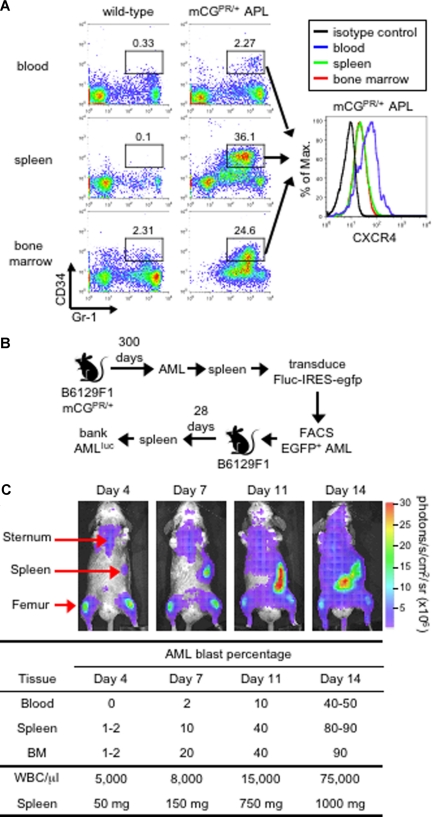
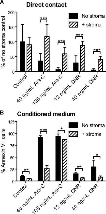
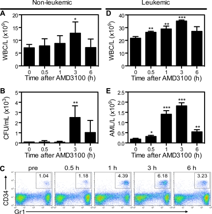
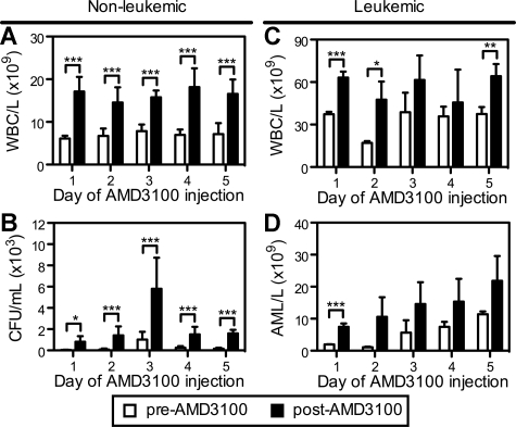
The CXCR4-SDF-1 axis plays a central role in the trafficking and retention of normal and malignant stem cells in the bone marrow (BM) microenvironment. Here, we used a mouse model of acute promyelocytic leukemia (APL) and a small molecule competitive antagonist of CXCR4, AMD3100, to examine the interaction of mouse APL cells with the BM microenvironment. APL cells from a murine cathepsin G-PML-RARalpha knockin mouse were genetically modified with firefly luciferase (APL(luc)) to allow tracking by bioluminescence imaging. Coculture of APL(luc) cells with M2-10B4 stromal cells protected the leukemia cells from chemotherapy-induced apoptosis in vitro. Upon injection into syngeneic recipients, APL(luc) cells rapidly migrated to the BM followed by egress to the spleen then to the peripheral blood with death due to leukostasis by day 15. Administration of AMD3100 to leukemic mice induced a 1.6-fold increase in total leukocytes and a 9-fold increase of circulating APL blast counts, which peak at 3 hours and return to baseline by 12 hours. Treatment of leukemic mice with chemotherapy plus AMD3100 resulted in decreased tumor burden and improved overall survival compared with mice treated with chemotherapy alone. These studies provide a proof-of-principle for directing therapy to the critical tethers that promote AML-niche interactions.


Figures

Figure 1
Figure 1
Murine APL model. (A) Immunophenotype of APL cells. Peripheral blood, splenocytes, and bone marrow cells from a healthy wild-type B6129F1 mouse and a leukemic murine cathepsin G-PML-RARα knock-in (mCGPR/+) B6129F1 mouse with APL were stained for the myeloid surface antigen Gr-1 and the early hematopoietic progenitor marker CD34. The level of CXCR4 expression on Gr-1+/CD34+ in mCGPR/+ APL cells is shown in the histogram along with the isotype control (black line). The indicated gates were used to determine the percentage of cells positive for Gr-1 and CD34. (B) Generation of luciferase-labeled acute myeloid leukemia (APLluc) cells. APL cells obtained from the spleen of a mCGPR/+ knock-in mouse were transduced with a bicistronic retroviral vector containing firefly luciferase upstream of EGFP (Fluc-IRES-egfp). Following transduction, EGFP+ APL cells were purified by fluorescence-activated cell sorting (FACS) and passaged in genetically compatible B6129FI recipients. These secondary recipients developed a rapidly fatal acute leukemia characterized by pronounced leukocytosis, anemia, thrombocytopenia, and massive hepatosplenomegaly with leukemic cell infiltration. APLluc cells obtained from the spleens of secondary recipients were frozen and banked 28 days after injection. (C) Kinetics of APLluc engraftment and expansion. Genetically compatible B6129F1 recipients were injected with 106 APLluc cells. Tumor trafficking and growth were assessed at various time intervals by bioluminescence imaging. Images of a representative mouse are shown. Photon flux is indicated in the color scale bar. WBC counts were determined by automated counting and the percentage of leukemic blasts in the blood, spleen, and bone marrow by flow cytometry.
Figure 2
Figure 2
In vitro sensitivity of APL cells to chemotherapy. (A) Decreased apoptosis of APLluc cells following culture with stromal cells. APLluc cells (103 cells/well) were cultured in 96-well black-walled tissue culture plates in the presence or absence of the murine bone marrow stromal cell line M2-10B4. After 48 hours, APLluc cells were incubated for an additional 48 hours in medium containing 40 ng/mL cytarabine (Ara-C), 105 ng/mL Ara-C, 12 ng/mL daunorubicin (DNR), 40 ng/mL DNR, or vehicle alone (control). APLluc cell survival was assessed by bioluminescent imaging and results are presented as a percent of the no stroma control. Each bar represents the mean plus or minus SD of 2 separate experiments, for which each sample was assayed in quadruplicate. (B) Decreased apoptosis of APL cells following culture in M2-10B4–conditioned medium. Cell-free culture supernatant was obtained from 3-day cultures of M2-10B4 stromal cells. APL cells (103 cells/well) were incubated for 48 hours in unconditioned medium (no stroma) or M2-10B4–conditioned medium (+stroma) containing 40 ng/mL Ara-C, 105 ng/mL Ara-C, 12 ng/mL DNR, 40 ng/mL DNR, or vehicle alone (control). APL cell death was assessed by flow cytometry using annexin V–PE and 7-amino-actinomycin D. Each bar represents the mean ± SD of 2 separate experiments, for which each sample was assayed in triplicate. *P < .05; **P < .01; and ***P < .001.
Figure 3
Figure 3
AMD3100 induces a rapid and transient mobilization of normal hematopoietic progenitor cells and APL blasts into the peripheral blood. Syngeneic B6129F1 recipient mice were left untreated (nonleukemic) or injected intravenously with APL cells (leukemic). Twelve days after APL injection, mice were treated with a single subcutaneous dose of 5 mg/kg AMD3100. Peripheral blood samples were collected immediately before and 0.5, 1, 3, and 6 hours after AMD3100 administration. (A) Total white blood cell (WBC) counts per microliter of peripheral blood in nonleukemic mice were determined by automated counting. (B) Colony-forming units in the peripheral blood of nonleukemic mice. (C) Representative flow cytometry profiles showing mobilization of Gr1+CD34+ APL blast cells following treatment with AMD3100. (D) Total white blood cell counts per microliter of peripheral blood in leukemic mice were determined by automated counting. (E) APL blast cell counts per microliter of peripheral blood in leukemic mice were determined by automated counting and flow cytometry of Gr1+CD34+ APL blast cells. Each bar represents the mean ± SD of a single experiment, for which each sample was assayed in quadruplicate. Results are representative of 3 separate experiments. *P < .05; **P < .01; and ***P < .001.
Figure 4
Figure 4
Repetitive mobilization of normal hematopoietic stem cells and APL blasts into the peripheral blood. Syngeneic B6129F1 recipient mice were left untreated (nonleukemic) or injected intravenously with 106 APL cells (leukemic). Twelve days after APL injection, mice were treated with a single subcutaneous dose of 5 mg/kg AMD3100 for 5 consecutive days. Peripheral blood samples were collected immediately before (pre-AMD3100) and 3 hours after (post-AMD3100) each daily dose of AMD3100. (A,C) Total white blood cell counts per microliter of peripheral blood in nonleukemic (A) and leukemic (C) mice. (C) Colony-forming units in the peripheral blood of nonleukemic mice. (D) Blast cell counts per microliter of peripheral blood in leukemic mice. APL blast cell counts per microliter of peripheral blood in leukemic mice were determined by automated counting and flow cytometry of Gr1+CD34+ APL blast cells. Each bar represents the mean ± SD of a single experiment, for which each sample was assayed in triplicate. *P < .05; **P < .01; and ***P < .001.
Figure 5
Figure 5
AMD3100 does not mobilize APL cells from the peritoneal cavity. (A) Kinetics of APL progression following intraperitoneal administration of APL cells. Syngeneic B6129F1 recipient mice (n = 10) were injected intraperitoneally with 106 APL cells and the percentage of Gr-1+CD34+ APL cells in the peritoneum, peripheral blood, spleen, and BM was determined weekly by flow cytometry. APL cells expanded into the peritoneal cavity during the first 2 weeks followed by engraftment in the BM and spleen during the third and fourth weeks. (B,C) Fifteen (B) or 22 (C) days after APL injection, leukemic mice were treated with a single subcutaneous dose of 5 mg/kg AMD3100 and killed 3 hours later. Baseline peritoneum and peripheral blood samples were collected immediately before (pre-AMD) administration of AMD3100. AMD3100-mobilized APL cells into the peripheral blood (B) during the fourth week, but not from the peritoneal cavity (A) during the second week.
Figure 6
Figure 6
AMD3100 sensitization of APL to Ara-C. (A-C) Syngeneic B6129F1 recipient mice (n = 29) were intravenously injected with 106 APLluc cells. Twelve days after APL injection, mice were left untreated (control; n = 6) or treated with AMD3100 alone (n = 7), Ara-C alone (n = 8), or the combination of AMD3100 and Ara-C (n = 8). Mice treated with chemotherapy received a single subcutaneous injection of Ara-C (500 mg/kg) on days 12 and 13 after APL injection. Mice treated with AMD3100 received subcutaneous injections of AMD3100 (5 mg/kg) 1 hour before and 3 hours after each Ara-C injection. (A) In vivo bioluminescent imaging of APLluc cells. One representative animal for each group is shown over time. Photon flux is indicated in the color scale bar. Red arrow indicates initiation of treatment with AMD3100 and/or Ara-C. (B) Expansion of APLluc cells was quantified in emitted photons over total body area (ventral view). BLI signal intensity at days 15, 19, and 23 after APL injection was significantly reduced in mice receiving the combination of AMD3100 and Ara-C compared with mice receiving Ara-C alone. Each bar represents the mean ± SD of a single experiment with the number of mice in each group exactly as described earlier in the legend. (C) Kaplan-Meier plot of overall survival of mice. Overall survival of leukemic mice is significantly prolonged when mice are treated with the combination of AMD3100 and Ara-C (P < .001 between Ara-C versus Ara-C + AMD3100 cohorts). (D) Syngeneic B6129F1 recipient mice (n = 30) were intravenously injected with 106 nontransduced APL cells. Twelve days after APL injection, mice were left untreated (control; n = 5) or treated with AMD3100 alone (n = 5), Ara-C alone (n = 10), or the combination of AMD3100 and Ara-C (n = 10) exactly as described earlier in the legend. Overall survival of leukemic mice is significantly prolonged when mice are treated with the combination of AMD3100 and Ara-C (P < .001 between Ara-C vs Ara-C + AMD3100 cohorts). *P < .05; **P < .01; and ***P < .001.

Comment in

  • Another nail in the AML coffin.
    Abboud CN. Abboud CN. Blood. 2009 Jun 11;113(24):6045-6. doi: 10.1182/blood-2009-03-189803. Blood. 2009. PMID: 19520813 No abstract available.
  • Priming reloaded?
    Heuser M, Kuchenbauer F, Argiropoulos B, Sekulovic S, Leung M, Stasiak M, Ganser A, Humphries RK. Heuser M, et al. Blood. 2009 Jul 23;114(4):925-6; author reply 926-7. doi: 10.1182/blood-2009-04-217299. Blood. 2009. PMID: 19628718 No abstract available.

References

    1. Nervi B, Link DC, DiPersio JF. Cytokines and hematopoietic stem cell mobilization. J Cell Biochem. 2006;99:690–705. - PubMed
    1. Ma Q, Jones D, Springer TA. The chemokine receptor CXCR4 is required for the retention of B lineage and granulocytic precursors within the bone marrow microenvironment. Immunity. 1999;10:463–471. - PubMed
    1. Nagasawa T, Hirota S, Tachibana K, et al. Defects of B-cell lymphopoiesis and bone-marrow myelopoiesis in mice lacking the CXC chemokine PBSF/SDF-1. Nature. 1996;382:635–638. - PubMed
    1. Aiuti A, Webb IJ, Bleul C, Springer T, Gutierrez-Ramos JC. The chemokine SDF-1 is a chemoattractant for human CD34+ hematopoietic progenitor cells and provides a new mechanism to explain the mobilization of CD34+ progenitors to peripheral blood. J Exp Med. 1997;185:111–120. - PMC - PubMed
    1. Semerad CL, Christopher MJ, Liu F, et al. G-CSF potently inhibits osteoblast activity and CXCL12 mRNA expression in the bone marrow. Blood. 2005;106:3020–3027. - PMC - PubMed

Publication types

MeSH terms