. 2009 Mar 5;113(10):2290-7.
doi: 10.1182/blood-2007-05-089193. Epub 2008 Dec 2.

Dysfunctional homologous recombination mediates genomic instability and progression in myeloma

Affiliations

Dysfunctional homologous recombination mediates genomic instability and progression in myeloma

Masood A Shammas et al. Blood. .

Abstract

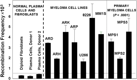
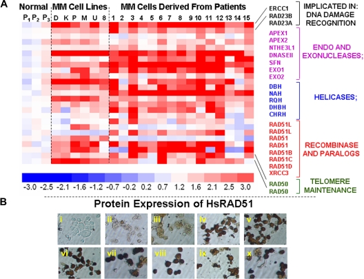
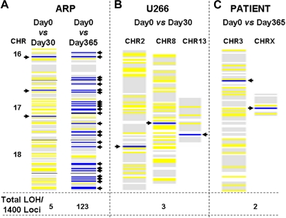
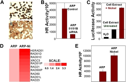
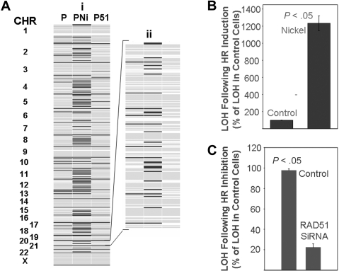
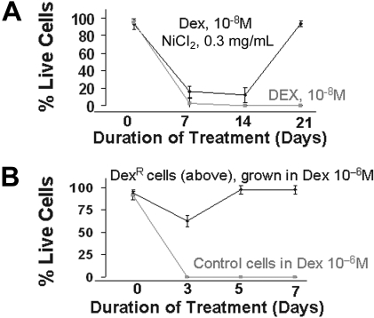
A prominent feature of most if not all cancers is a striking genetic instability, leading to ongoing accrual of mutational changes, some of which underlie tumor progression, including acquisition of invasiveness, drug resistance, and metastasis. Thus, the molecular basis for the generation of this genetic diversity in cancer cells has important implications in understanding cancer progression. Here we report that homologous recombination (HR) activity is elevated in multiple myeloma (MM) cells and leads to an increased rate of mutation and progressive accumulation of genetic variation over time. We demonstrate that the inhibition of HR activity in MM cells by small inhibitory RNA (siRNAs) targeting recombinase leads to significant reduction in the acquisition of new genetic changes in the genome and, conversely, the induction of HR activity leads to significant elevation in the number of new mutations over time and development of drug resistance in MM cells. These data identify dysregulated HR activity as a key mediator of DNA instability and progression of MM, with potential as a therapeutic target.


Figures

Figure 1
Figure 1
Increased HR activity in MM cells. HR activity was measured with the recombination plasmid substrate. Generation of the neomycin resistance as a measure of recombination activity was evaluated by counting neomycin-resistant colonies and total plasmid-transfected was evaluated by ampicillin-resistant colonies. The recombination frequency was calculated from the ratio of these 2 counts. Recombination frequency is presented as number of recombination events per million transfected plasmid copies.
Figure 2
Figure 2
Elevated expression of HR-related genes in MM cells. (A) Gene expression profile was measured in 3 normal plasma cell isolates (P1, P2, P3), 6 myeloma cell lines (D indicates ARD; K, ARK; P, ARP; M, MM1s; U, U266; and 8, RPMI8226), and 15 purified myeloma cells from patient bone marrows (1-15) with the use of HG-U133 gene arrays. The color scale at the bottom of the figure represents fold change in recombination- and repair-associated genes in myeloma cells relative to average expression in 3 normal plasma cell samples. (B) Protein levels of HsRAD51 and its paralogs were detected by immunostaining, with either a polyclonal antibody against HsRAD51, also recognizing RAD51B and RAD51C or monoclonal antibodies recognizing HsRAD51 or its paralogs individually. (i) Normal plasma cells, no primary antibody control; (ii) normal plasma cells (donor 1), HsRAD51/51B/51C; (iii) normal plasma cells (donor 2), HsRAD51/51B/51C; (iv) ARP myeloma cell line, HsRAD51/51B/51C; (v) MM1S myeloma cell line, HsRAD51/51B/51C; (vi) U266 myeloma cell line, HsRAD51/51B/51C; (vii) patient myeloma cells, HsRAD51; (viii) patient myeloma cells HsRAD51B; (ix) patient myeloma cells, HsRAD51C; and (x) patient myeloma cells, HsRAD51D.
Figure 3
Figure 3
Acquisition of new genomic changes in multiple myeloma cells over time. Myeloma cells were cultured for various durations. Aliquots of cells were removed at specified intervals and genomic DNA was isolated and evaluated for LOH with the use of HuSNP chips (Affymetrix). DNA from the cells harvested and frozen at day 0 was used to define the allelotype baseline, departures from which identified new LOH loci in cells harvested at later time points. Blue lines show new LOH loci in the regions of genome indicated, whereas yellow lines represent retention of preexisting LOH sites. The numbers at the bottom of each figure indicate total number of new LOH loci throughout genome, per 1400 sites investigated. (A) New LOH loci in ARP myeloma cells cultured for either 1 or 12 months, in the region of genome spanning chromosomes 16, 17, and 18, are shown as an example. (B) New LOH loci in U266 myeloma cells cultured for 1 month. (C) New LOH loci in genomic DNA samples, derived from a patient at 1-year intervals.
Figure 4
Figure 4
HR activity in myeloma cells is induced by nickel chloride and inhibited by siRNAs targeting recombinases. (A) Loss of RAD51 protein expression by siRNAs. Immunostaining using monoclonal antibody to HsRAD51 of ARP cells transfected with (I) control siRNA and (II) HsRAD51-specific siRNAs. (B) Loss of HR activity by siRNAs. HR activity was assessed using the plasmid substrate after the transfection of ARP cells with siRNA directed at HsRAD51 and RAD51B. Recombination frequency is presented as number of recombination events per million transfected plasmid copies. ARP control, MM cells transfected with control siRNAs; ARP 51/51B SiRNA, MM cells transfected with siRNAs directed at RAD51 ands RAD51B. (C) Effect of nickel on promoter regulation of HsRAD51. HsRAD51 promoter was cloned upstream of a luciferase gene in a mammalian expression vector. The resulting construct HsRAD51P-LUC was transfected into normal diploid fibroblasts and the cells were then exposed to nickel chloride (0.3 mg/mL). After a 2-hour exposure, cells were lysed and luciferase activity was assayed with a Luciferase Assay Kit (Clontech). Lanes show luciferase activity in water (lane 1), in ARP cells transfected with RAD51P-Luc (lane 2), and in ARP cells transfected with RAD51P-Luc and exposed to nickel chloride (lane 3). (D) Induction of HsRAD51 and related genes by nickel chloride in ARP cells. ARP myeloma cells, untreated (ARP) or treated with nickel chloride (0.3 mg/mL) for 24 hours (ARP-Ni) were harvested, total RNA was isolated, and processed for gene expression analysis using U133 arrays (Affymetrix). Gene expression is shown by intensity of red color. Color scale shows fold change of gene expression in treated cells relative to untreated ARP cells. (E) HR activity in untreated ARP cells (ARP) or ARP cells exposed to nickel chloride (ARP Nickel; 0.3 mg/mL) for 5 days.
Figure 5
Figure 5
Effect of homologous recombination on rate of mutation in myeloma cells. (A) ARP myeloma cells were either transfected every 2 weeks with control siRNAs for 90 days (lane 1), exposed to nickel chloride (0.3 mg/mL) for 5 days (lane 2), or transfected every 2 weeks with siRNAs targeting HsRAD51 for 90 days (lane 3). Genomic DNA was isolated and evaluated with huSNP arrays (Affymetrix) as described. A representative figure showing all chromosomes (i) and an enlarged version of chromosome 20 (ii) is shown as an example. Dark lines indicate new LOH loci. (B,C) Summary of change in LOH over all chromosomes; error bars indicate standard error of the mean of triplicate assays. (B) Induction of LOH in ARP cells after exposure to nickel chloride as a percentage of control cell values. (C) Reduction in LOH in ARP cells after suppression of HR by HsRAD51 directed siRNA as a percentage of control cell values.
Figure 6
Figure 6
Induction of drug resistance by elevated HR activity. (A) ARP myeloma cells were cultured in the presence of dexamethasone (10−8 mol/L) alone or dex (10−8 mol/L) and nickel chloride (0.3 mg/mL). Live cell number was determined weekly by trypan blue staining. After 2 weeks of culture, nickel was removed from the dexamethasone and nickel-treated cultures. The figure is representative of 3 independent experiments. (B) Dexamethasone-resistant MM cells obtained in panel A were cultured in higher concentrations of dex (10−6 mol/L). Live cell number of control and dexamethasone-resistant ARP cells was determined by trypan blue staining.

References

    1. Lengauer C, Kinzler KW, Vogelstein B. Genetic instabilities in human cancers. Nature. 1998;396:643–649. - PubMed
    1. Shmookler Reis RJ, Goldstein S. Loss of reiterated DNA sequences during serial passage of human diploid fibroblasts. Cell. 1980;21:739–749. - PubMed
    1. Srivastava A, Norris JS, Shmookler Reis RJ, Goldstein S. c-Ha-ras-1 proto-oncogene amplification and overexpression during the limited replicative life span of normal human fibroblasts. J Biol Chem. 1985;260:6404–6409. - PubMed
    1. Tlsty TD. Normal diploid human and rodent cells lack a detectable frequency of gene amplification. Proc Natl Acad Sci U S A. 1990;87:3132–3136. - PMC - PubMed
    1. Sweezy MA, Fishel R. Multiple pathways leading to genomic instability and tumorigenesis. Ann N Y Acad Sci. 1994;726:165–177. - PubMed

Publication types