

Lower orbital frontal white matter integrity in adolescents with bipolar I disorder
- PMID: 19050654
- PMCID: PMC2747245
- DOI: 10.1097/CHI.0b013e3181900421
Lower orbital frontal white matter integrity in adolescents with bipolar I disorder
Abstract
Objective: To examine white matter microstructure, as assessed via diffusion tensor imaging (DTI), in adolescents with bipolar I disorder compared with control volunteers.
Method: Twenty-six (12 male and 14 female subjects) adolescents (mean age, 16.0 years) with bipolar I disorder and 26 (14 male and 12 female subjects) control volunteers (mean age, 15.3 years) completed structural and DTI examinations. Fractional anisotropy (FA) and apparent diffusion coefficient (ADC) maps were compared between groups in the brain white matter using a voxelwise analysis after intersubject registration to Talairach space. Exploratory analyses were performed to assess structure-function correlations in a subgroup of 11 patients with available neuropsychological measures.
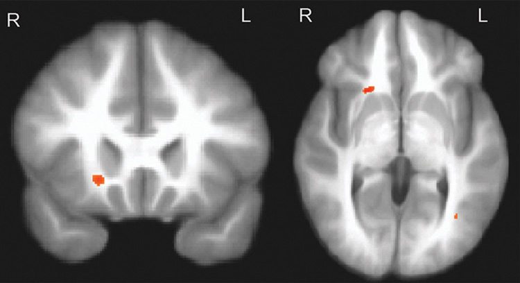
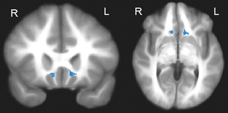
Results: Compared with the control volunteers, the patients demonstrated abnormalities in white matter regions predicted to differ a priori between groups, including lower FA in the right orbital frontal lobe and higher ADC in the right and left subgenual region (p <.005, uncorrected; cluster size >or= 100). There were no areas of higher FA or lower ADC in patients compared with control volunteers. Lower FA across regions that differed significantly between groups correlated significantly with slower visuomotor speed among patients with bipolar disorder.
Conclusions: Abnormalities involving the orbital frontal and subgenual white matter in adolescents with bipolar disorder are consistent with neurobiological models that implicate dysregulation of affective systems and impulsivity in the pathophysiology of the disorder. Preliminary findings suggest that white matter abnormalities in pediatric bipolar disorder have functional correlates and may be useful in constructing neurobiological models of the disorder.
Conflict of interest statement
Disclosure: The authors report no conflicts of interest.
Figures
References
-
- Aylward EH, Roberts-Twillie JV, Barta PE, et al. Basal ganglia volumes and white matter hyperintensities in patients with bipolar disorder. Am J Psychiatry. 1994;151:687–693. - PubMed
-
- Ahn KH, Lyoo IK, Lee HK, et al. White matter hyperintensities in subjects with bipolar disorder. Psychiatry Clin Neurosci. 2004;58:516–521. - PubMed
-
- Kieseppä T, van Erp TG, Haukka J, et al. Reduced left hemispheric white matter volume in twins with bipolar I disorder. Biol Psychiatry. 2003;54:896–905. - PubMed
-
- Strakowski SM, Wilson DR, Tohen M, et al. Structural brain abnormalities in first-episode mania. Biol Psychiatry. 1993;33:602–609. - PubMed
-
- Tkachev D, Mimmack ML, Ryan MM, et al. Oligodendrocyte dys-function in schizophrenia and bipolar disorder. Lancet. 2003;362:798–805. - PubMed
