. 2009 Feb;17(2):334-42.
doi: 10.1038/mt.2008.256. Epub 2008 Dec 2.

Delivery of therapeutic proteins as secretable TAT fusion products

Affiliations

Delivery of therapeutic proteins as secretable TAT fusion products

Marcella Flinterman et al. Mol Ther. 2009 Feb.

Abstract

The trans-acting activator of transcription (TAT) protein transduction domain (PTD) mediates the transduction of peptides and proteins into target cells. The TAT-PTD has an important potential as a tool for the delivery of therapeutic agents. The production of TAT fusion proteins in bacteria, however, is problematic because of protein insolubility and the absence of eukaryotic post-translational modification. An attractive alternative, both for in vitro protein production and for in vivo applications, is the use of higher eukaryotic cells for secretion of TAT fusion proteins. However, the ubiquitous expression of furin endoprotease (PACE or SPC1) in the Golgi/endoplasmic reticulum, and the presence of furin recognition sequences within TAT-PTD, results in the cleavage and loss of the TAT-PTD domain during its secretory transition through the endoplasmic reticulum and Golgi. In this study, we show the development of a synthetic TATkappa-PTD in which mutation of the furin recognition sequences, but retention of protein transduction activity, allows secretion of recombinant proteins, followed by successful uptake of the modified protein, by the target cells. This system was used to successfully secrete marker protein, green fluorescent protein (GFP), and apoptin, a protein with tumor-specific cytotoxicity. Detection of GFP, phosphorylation, and induction of cell death by TATkappa-GFP-apoptin indicated that the secreted proteins were functional in target cells. This novel strategy therefore has important potential for the efficient delivery of therapeutic proteins.


Figures

<b>Figure 1</b>
Figure 1
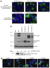
Transient transfection of 293T cells with pSec constructs. (a) 293T cells were transfected with different pSec constructs; 48 hours after transfection cells were fixed, permeabilized, counterstained with 4′,6-diamino-2-phenylindole–containing mounting solution and green fluorescent protein (GFP) expression was examined by fluorescence microscopy. Scale bar indicates 10 µm. (b) Secretion in medium after transient transfection of 293T cells with pSec constructs. Supernatant from each well was collected at the indicated time points, lysed and GFP was detected by western blot analysis using primary polyclonal antibody to GFP (Abcam) and secondary horseradish peroxidase–linked antibody to rabbit (GE healthcare). TAT, trans-acting activator of transcription.
<b>Figure 2</b>
Figure 2
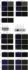
Green fluorescent protein (GFP) expression in 293T/TAT-GFP and TATκ-GFP stable clones. (a) The protein sequence of trans-acting activator of transcription (TAT) and TATκ are indicated. 293T cells were stably transfected with TAT-GFP or TATκ-GFP as indicated in the Materials and Methods and clones were picked after 3 weeks. Clones were fixed, permeabilized, counterstained with 4′,6-diamino-2-phenylindole–containing mounting solution and GFP expression was examined by fluorescence microscopy. (b) Secretion in medium from 293T TAT-GFP and TATκ-GFP clones. Supernatant from each well was collected 24 hours after plating, and GFP was detected by western blot analysis. (c) 293T/TATκ-GFP/clone 2 (c2) treated with brefeldin A. 293T/TATκ-GFP/c2 was seeded in a 8-well chamber well slide at a concentration of 100,000 cells per well and treated the following day with different concentrations brefeldin A. Supernatant was collected after 3 hours, centrifuged to remove any floating cells, lysed and GFP expression was examined by western blot analysis. (d) Twenty-four hours after brefeldin A treatment cells were fixed, permeabilized and GFP expression was examined by fluorescence microscopy. All scale bars indicate 10 µm. HIV, human immunodeficiency virus.
<b>Figure 3</b>
Figure 3
Target cells are efficiently transduced by TATκ-GFP. (a) H357 and Saos-2 cells were treated with supernatant from 293T/TATκ-GFP/c2 or 293T. After 30 minutes, cells were fixed, permeabilized, and examined for green fluorescent protein (GFP) expression. (b) GFP expression in target cells detected by western blot analysis. Saos-2 or H357 cells treated for 30 minutes with supernatant of 293T cells or 293T/TATκ-GFP/c2 and lysed for western blot analysis of GFP expression. Supernatant of 293T/TATκ-GFP/c2 was used as positive control for GFP. (c) Western blot analysis showing the presence of GFP in concentrated and nonconcentrated supernatant. (d) Saos-2 and (e) HeLa cells were treated for 30 minutes with concentrated supernatant from 293T or 293T/TATκ-GFP/c2, cells were fixed, permeabilized, and analyzed for GFP expression by fluorescence microscopy. (f) Fixed and unfixed transduced target cells. Saos-2 cells were treated with concentrated supernatant from 293T/TATκ-GFP/c2 and 293T cells for 30 minutes and GFP expression was either examined by fluorescence microscopy immediately or after fixation with 2% paraformaldehyde and permeabilization with 0.2% Triton X-100. All scale bars indicate 40 µm. FCS, fetal calf serum; TAT, trans-acting activator of transcription.
<b>Figure 4</b>
Figure 4
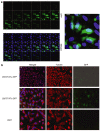
Green fluorescent protein (GFP) is localized inside target cells. (a) Confocal microscopy showing GFP expression in target cells. Saos-2 cells were treated with concentrated supernatant from 293T/TATκ-GFP/c2 and 293T cells for 30 minutes and examined by confocal microscopy. The upper left panel shows a Z-stack of GFP expression, the lower left panel of the merged image [showing both 4′,6-diamino-2-phenylindole (DAPI) and GFP] and right panel the complete image, stack size 0.3 µm. (b) Colocalization of tubulin and GFP in Saos-2 cells transduced with TATκ-GFP. Target cells were treated with concentrated or nonconcentrated supernatant for 30 minutes and cells were fixed, permeabilized, blocked, and stained for tubulin expression using a primary mouse antibody to tubulin (Sigma, Poole, UK) and a secondary antibody to mouse labeled with Texas Red. Cells were counterstained with DAPI-containing mounting solution and were examined for GFP and tubulin expression by fluorescence microscopy. All scale bars indicate 10 µm, except for top image in b, where it indicates 40 µm. TAT, trans-acting activator of transcription.
<b>Figure 5</b>
Figure 5
Green fluorescent protein (GFP) uptake by target cells cocultured in transwells with 293T/TATκ-GFP/c2. FDCP1 cells were cocultured for 3 days in transwells together with either 293T/TATκ-GFP/c2 or 293T cells. (a) Western blot showing TATk-GFP level in supernatant in transwell. Supernatant was collected after 3 days of coculture and centrifuged for 5 minutes to remove FDCP1 cells. (b) GFP uptake in FDCP1 cells after 3 days of coculture was shown by FACSSCAN. TAT, trans-acting activator of transcription.
<b>Figure 6</b>
Figure 6
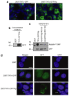
Expression and secretion 293T/TATκ-GFP/Ap/c1. (a) 293T cells were stably transfected with TATκ-GFP/Ap as indicated in Materials and Methods and after 72 hours cells were put under selection with 400 µg/ml zeocin. Cells from 293T/TATκ-GFP/Ap/c1 were fixed, permeabilized, and examined for TATκ-GFP/Ap expression by fluorescence microscopy. 293T/TATκ-GFP/c2 was used as a control. (b) Supernatant collected from 293T cells and 293T/TATκ-GFP/Ap/c1 was concentrated, lysed, and examined for TATκ-GFP/Ap secretion by western blot analysis. (c) Green fluorescent protein (GFP)-apoptin is phosphorylated in 293T/TATκ-GFP/Ap/c1. Cells were lysed and phosphorylation of apoptin was detected using the apoptin phosphospecific antibody VP3T108P, as positive control for apoptin phosphorylation SV40 transformed human fibroblasts 1BR3-N infected with adenovirus expressing GFP-apoptin (Ad-GFP/Ap) at an MOI 50 and 100 were used. (d) Saos-2 cells were treated for 30 minutes with concentrated supernatant from 293T, 293T/TATκ-GFP/c2, or 293T/TATκ-GFP/Ap/c1, cells were fixed, permeabilized, and analyzed for GFP or GFP-apoptin expression by fluorescence microscopy. All scale bars indicate 10 µm, except for left image in a, where it indicates 40 µm. DAPI, 4′,6-diamino-2-phenylindole; MOI, multiplicity of infection; TAT, trans-acting activator of transcription.

References

    1. Hacein-Bey-Abina S, Von Kalle C, Schmidt M, McCormack MP, Wulffraat N, Leboulch P, et al. LMO2-associated clonal T cell proliferation in two patients after gene therapy for SCID-X1. Science. 2003;302:415–419. - PubMed
    1. Mae M., and , Langel U. Cell-penetrating peptides as vectors for peptide, protein and oligonucleotide delivery. Curr Opin Pharmacol. 2006;6:509–514. - PubMed
    1. Wadia JS., and , Dowdy SF. Transmembrane delivery of protein and peptide drugs by TAT-mediated transduction in the treatment of cancer. Adv Drug Deliv Rev. 2005;57:579–596. - PubMed
    1. Guelen L, Paterson H, Gäken J, Meyers M, Farzaneh F., and , Tavassoli M. TAT-apoptin is efficiently delivered and induces apoptosis in cancer cells. Oncogene. 2004;23:1153–1165. - PubMed
    1. Zhuang SM, Shvarts A, Jochemsen AG, van Oorschot AA, van der Eb AJ., and , Noteborn MH. Differential sensitivity to Ad5 E1B-21kD and Bcl-2 proteins of apoptin-induced versus p53-induced apoptosis. Carcinogenesis. 1995;16:2939–2944. - PubMed

Publication types

MeSH terms

Substances

LinkOut - more resources