Multicenter Study
. 2009 May;17(5):656-63.
doi: 10.1038/ejhg.2008.226. Epub 2008 Dec 3.

In-frame deletion in the seventh immunoglobulin-like repeat of filamin C in a family with myofibrillar myopathy

Affiliations
Multicenter Study

In-frame deletion in the seventh immunoglobulin-like repeat of filamin C in a family with myofibrillar myopathy

Alexey Shatunov et al. Eur J Hum Genet. 2009 May.

Abstract

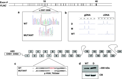
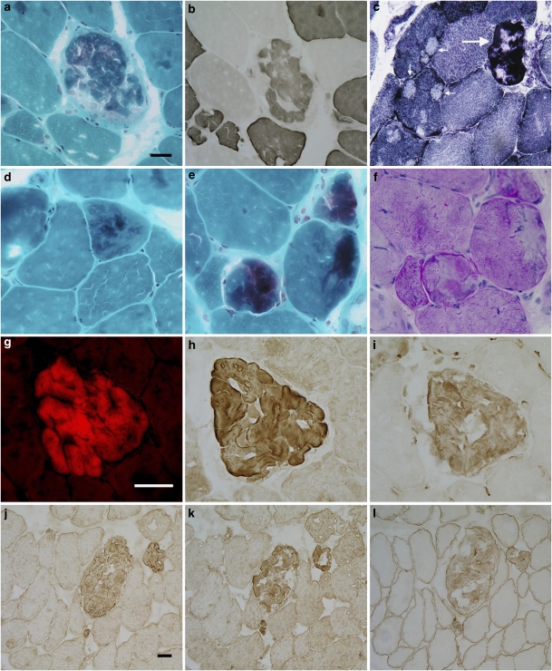
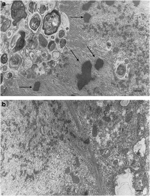
Myofibrillar myopathies (MFMs) are an expanding and increasingly recognized group of neuromuscular disorders caused by mutations in DES, CRYAB, MYOT, and ZASP. The latest gene to be associated with MFM was FLNC; a p.W2710X mutation in the 24th immunoglobulin-like repeat of filamin C was shown to be the cause of a distinct type of MFM in several German families. We studied an International cohort of 46 patients from 39 families with clinically and myopathologically confirmed MFM, in which DES, CRYAB, MYOT, and ZASP mutations have been excluded. In patients from an unrelated family a 12-nucleotide deletion (c.2997_3008del) in FLNC resulting in a predicted in-frame four-residue deletion (p.Val930_Thr933del) in the seventh repeat of filamin C was identified. Both affected family members, mother and daughter, but not unrelated control individuals, carried the p.Val930_Thr933del mutation. The mutation is transcribed and, based on myopathological features and immunoblot analysis, it leads to an accumulation of dysfunctional filamin C in the myocytes. The study results suggest that the novel p.Val930_Thr933del mutation in filamin C is the cause of MFM but also indicate that filamin C mutations are a comparatively rare cause of MFM.


Figures

Figure 1
Figure 1
Molecular characterization of the identified FLNC mutation. Schematic drawing of the FLNC gene and filamin C protein (bottom part) showing the molecular effects of the FLNC deletion. (a) cDNA sequence alignment of a fragment of FLNC exon 18; the underlined 12 nucleotides (c.2997–3008) are deleted from the patient's sequence. (b) GeneScan profiles show a 12-bp difference in PCR-produced fragments amplified from the affected daughter's (D) and her mother's (M) genomic DNA (gDNA), compared with a single peak in the control (WT). Two transcript species with a 12-bp size difference were also identified in the biopsy muscle tissue obtained from the affected daughter, but only the longer transcript was present in the control sample. (c) The 12-nucleotide deletion in exon 18 is predicted to result in an in-frame deletion of four amino acids VKYT (p.Val930_Thr933del) within the seventh repeat of filamin C. (d) Immunoblot analysis of the muscle tissue proteins obtained from the patient (D) revealed a slight increase in the quantity of filamin C when compared with the control (WT). Myosin band of 205 kDa stained with Coomassie Brilliant Blue R (CB) shows equal protein loading. ABD, actin-binding domain; CHD 1 and 2, calponin homology domains.
Figure 2
Figure 2
Histochemical and immunohistochemical findings in a patient with filaminopathy. (a) A large fiber containing a hyaline inclusion. (b) ATPase and (c) oxidative activities are partially reduced in the abnormal fiber area. Some adjacent fibers show several core-like lesions (arrows in c). Other types of lesions: (d) a thin, vermiform inclusion, (e) three muscle fibers containing demarcated abnormal areas, and (f) displaying some PAS positivity. Serial consecutive sections show an inclusion displaying congophilia (g), filamin C (h), ubiquitin (i), desmin (j), myotilin (k), and dystrophin (l) immunoreactivity. (a, d, and e) modified trichrome; (b) ATPase 4.35; (c) NADH; (f) PAS; (g) Congo Red; (h) filamin C; (i) ubiquitin; (j) desmin; (k) myotilin; and (l) dystrophin staining. Bar in a=40 μm (refers to a through f); bar in g=50 μm (refers to g through i); bar in j=25 μm (refers to j through l).
Figure 3
Figure 3
Ultrastructural analysis of skeletal muscle. (a) Widespread myofibrillary abnormalities, abundant rod formation (arrows), granulofilamentous material, and filamentous profiles. A large autophagic vacuole containing myeloid structures and membranous debris. (b) Remnants of filaments, Z-disc material, and foci of mitochondria. Bar=0.2 μm.

References

    1. Nakano S, Engel AG, Waclawik AJ, et al. Myofibrillar myopathy with abnormal foci of desmin positivity. 1. Light and electron microscopy analysis of 10 cases. J Neuropathol Exp Neurol. 1996:55, 549–562. - PubMed
    1. Engel AG. Myofibrillar myopathy. Ann Neurol. 1999;46:681–683. - PubMed
    1. Selcen D, Ohno K, Engel AG. Myofibrillar myopathy: clinical, morphological and genetic studies in 63 patients. Brain. 2004;127:439–451. - PubMed
    1. Vicart P, Caron A, Guicheney P, et al. A missense mutation in the alphaB-crystallin chaperone gene causes a desmin-related myopathy. Nat Genet. 1998;20:92–95. - PubMed
    1. Selcen D, Engel AG.Myofibrillar myopathy GeneReviews , ( http://www.genereviews.org/ ) 2008

Publication types

MeSH terms