Comparative Study
. 2009 Jan;292(1):94-106.
doi: 10.1002/ar.20768.

Characterization and classification of zebrafish brain morphology mutants

Affiliations
Comparative Study

Characterization and classification of zebrafish brain morphology mutants

Laura Anne Lowery et al. Anat Rec (Hoboken). 2009 Jan.

Abstract

The mechanisms by which the vertebrate brain achieves its three-dimensional structure are clearly complex, requiring the functions of many genes. Using the zebrafish as a model, we have begun to define genes required for brain morphogenesis, including brain ventricle formation, by studying 16 mutants previously identified as having embryonic brain morphology defects. We report the phenotypic characterization of these mutants at several timepoints, using brain ventricle dye injection, imaging, and immunohistochemistry with neuronal markers. Most of these mutants display early phenotypes, affecting initial brain shaping, whereas others show later phenotypes, affecting brain ventricle expansion. In the early phenotype group, we further define four phenotypic classes and corresponding functions required for brain morphogenesis. Although we did not use known genotypes for this classification, basing it solely on phenotypes, many mutants with defects in functionally related genes clustered in a single class. In particular, Class 1 mutants show midline separation defects, corresponding to epithelial junction defects; Class 2 mutants show reduced brain ventricle size; Class 3 mutants show midbrain-hindbrain abnormalities, corresponding to basement membrane defects; and Class 4 mutants show absence of ventricle lumen inflation, corresponding to defective ion pumping. Later brain ventricle expansion requires the extracellular matrix, cardiovascular circulation, and transcription/splicing-dependent events. We suggest that these mutants define processes likely to be used during brain morphogenesis throughout the vertebrates. Anat Rec, 2009. (c) 2008 Wiley-Liss, Inc.


Figures

Figure 1
Figure 1
Brain ventricle injections of midline separation defects mutants (Class 1, described in the text). Dorsal views of living, anesthetized embryos are shown, anterior to right, at 22 hpf (A–C,G–I) and 32–36 hpf (D–F,J–L) with brightfield microscopy. Ventricles are injected with Rhodamine-dextran. Compared to WT (A, D), the left and right sides of the brain tube do not open uniformly in the midline separation mutants (B–L). In nok (B,E) dye injected into the hindbrain ventricle does not move. In the other mutants, (C,F) ome, (G,J) has, (H,K) zon, (I,L) atl, there are regions where the tube opens separated by places where the sides appear to be touching (arrows). The ventricles of WT are labeled for comparison. F: forebrain ventricle, M: midbrain ventricle, H: hindbrain ventricle.
Figure 2
Figure 2
Brain morphology comparison of Class 1 and Class 2. (A–C) Hindbrain dorsal views of living, anesthetized embryos are shown, anterior to right, at 22 hpf with brightfield microscopy, after dye injection. While Class 1 has locations along the brain tube where the left and right sides are opposed (B, has mutant), the sides of Class 2 brain tube separate normally (C, lnf mutant). (D–F) Hindbrain horizontal confocal sections after soaking embryos in BODIPY-ceramide to highlight cell outlines. Bright green identified hindbrain ventricle space. Neuroepithelial cells of Class 1 touch at the midline, or are perhaps fused (E, arrows, zon mutant), however fluid separates left and right sides of Class 2 (F, lnf mutant).
Figure 3
Figure 3
Epithelial junction analysis of ome and has mutants. Confocal images of 24 hpf flat-mounted embryos in horizontal section through midbrain and hindbrain. (A–C) Phalloidin-Texas Red labels adherens junction-associated actin of wild type (A), ome mutant (B), has mutant (C). Actin is enriched at the apically localized adherens-junctions, and actin localization is normal in the mutants. (D–F) Mpp5 antibody labeling (green) of wild type (D) ome mutant (E), and has mutant (F) with phalloidin-Texas Red as counterstain (red). Mpp5 is apically-localized in wild-type. In ome, while some Mpp5 localizes normally to the junctions, it is also present throughout the entire neuroepithelium (E). Localization is normal in has (F). Part of the midbrain ventricular surface is not visible in the plane of section, but junctions are normal in those planes. M midbrain, H hindbrain.
Figure 4
Figure 4
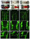
Brain ventricle injections and antibody labelings for reduced ventricle size Class 2. Dorsal views of living, anesthetized embryos are shown, anterior to right, at 22 hpf (A–F) and 36 hpf (G–L) with brightfield microscopy. Ventricles are injected with Rhodamine-dextran. The brain ventricles of lnf (B,H), ful (C,I), ott (D,J), log (E,K), and esa (F,L) are all similarly reduced compared to wild-type (A,G). (M–R) Dorsal views of 36 hpf hindbrain flatmounts, anterior is to the top, after labeling with the RMO44 Ab (reticulospinal neurons) shows reduced number of cell bodies and axons in lnf (N), ful (O), ott (P), and log (Q), compared to wild-type (M), although esa (R) appears similar to wild-type (M). (S–X) Dorsal views of 30 hpf hindbrain flatmounts, anterior is to the top, after labeling with the zn8 Ab (hindbrain commissural neurons) shows various levels of reduced commissures in lnf (T), ful (U), ott (V), and log (W), although esa (X) is indistinguishable from wild-type (S). (Y,Z) Lateral views of 36 hpf forebrain and midbrain flatmounts, anterior is to the left, dorsal is to the top, after labeling with acetylated tubulin Ab, which identifies the early axon scaffolds, shows that esa axonal pathfinding is severely disrupted, with the axons having a “feathered” appearance rather than fasciculating normally (Z), compared to wild-type (Y).
Figure 5
Figure 5
Brain ventricle injections and neuronal antibody labelings for MHB abnormalities Class 3. Dorsal views of living, anesthetized embryos are shown, anterior to right, at 22 hpf (A–C) and 36 hpf (D–F) with brightfield microscopy. Ventricles are injected with Rhodamine-dextran. At 22 hpf, both sly and gup (B,C) show an abnormal midbrain-hindbrain boundary. By 36 hpf, the sly and gup (E,F) boundary region has mostly recovered compared to WT (D), although the forebrain and midbrain ventricles are not as large as in WT. The ventricles of WT are labeled for comparison. F: forebrain ventricle, M: midbrain ventricle, H: hindbrain ventricle. (G–I) Dorsal views of 36 hpf hindbrain flatmounts, anterior is to the top, after labeling with the RMO44 Ab (reticulospinal neurons) shows disruption in axon pathfinding in both sly (H) and gup (I), compared to wild-type (G). (J–L) Dorsal views of 30 hpf hindbrain flatmounts, anterior is to the top, after labeling with the zn8 Ab (hindbrain commissural neurons) shows reduced commissures and disruption in axon pathfinding in both sly (H) and gup (I), compared to wild-type (G). (M–O) Lateral views of 36 hpf forebrain and midbrain flatmounts, anterior is to the left, dorsal is to the top, after labeling with acetylated tubulin Ab, which identifies the early axon scaffolds, shows that sly axonal pathfinding is disrupted (N), although gup (O) looks similar to wild-type (M).
Figure 6
Figure 6
Brightfield microscopy images of absence of lumen inflation mutant Class 4. Dorsal views of living, anesthetized embryos are shown, anterior to right. While the snk mutant at 22 hpf (B) has no visible ventricles and thus ventricles are not injected with dye, by 30 hpf (D), there are small ventricles in which dye can be injected, showing smaller but relatively normal shaping. F: forebrain ventricle, M: midbrain ventricle, H: hindbrain ventricle.
Figure 7
Figure 7
Later brain ventricle expansion class. Lateral views of living, anesthetized embryos are shown, anterior to right, at 28 hpf, with brightfield microscopy. The vip mutant shows reduced hindbrain ventricle height (B, red bracket) compared to wild-type (A). The nat mutant shows more severe brain ventricle height reduction (C, red bracket). The wis mutant also shows significantly reduced hindbrain ventricle height (D, red bracket), in addition to reduced pigmentation and other brain morphology abnormalities not shown in this figure.
Figure 8
Figure 8
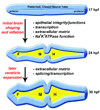
Gene functions required for early brain morphogenesis. Processes involved in initial brain shaping and inflation include midline separation (requiring epithelial integrity/junctions), other mechanisms affecting brain morphology (requiring transcription, among other unknown factors), midbrain-hindbrain boundary formation (requiring extracellular matrix), and brain lumen inflation (requiring Na+ K+ ATPase activity). Later brain ventricle expansion requires the extracellular matrix in order to maintain normal ventricle height, as well as splicing/transcription, which contributes to normal brain morphology. F:forebrain ventricle, M:midbrain ventricle, H:hindbrain ventricle.

References

    1. Amsterdam A, Nissen RM, Sun Z, Swindell EC, Farrington S, Hopkins N. Identification of 315 genes essential for early zebrafish development. Proc Natl Acad Sci U S A. 2004;101:12792–12797. - PMC - PubMed
    1. Antonova E, Sharma T, Morris R, Kumari V. The relationship between brain structure and neurocognition in schizophrenia: a selective review. Schizophr Res. 2004;70:117–145. - PubMed
    1. Bayer SA, Altman J. The human brain during the early first trimester. Boca Raton: CRC Press; 2007.
    1. Biehlmaier O, Makhankov Y, Neuhauss SC. Impaired retinal differentiation and maintenance in zebrafish laminin mutants. Invest Ophthalmol Vis Sci. 2007;48:2887–2894. - PubMed
    1. Bingham S, Chaudhari S, Vanderlaan G, Itoh M, Chitnis A, Chandrasekhar A. Neurogenic phenotype of mind bomb mutants leads to severe patterning defects in the zebrafish hindbrain. Dev Dyn. 2003;228:451–463. - PMC - PubMed

Publication types

LinkOut - more resources