. 2009 Feb;83(4):1962-8.
doi: 10.1128/JVI.01271-08. Epub 2008 Dec 3.

Differential sensitivity of differentiated epithelial cells to respiratory viruses reveals different viral strategies of host infection

Affiliations

Differential sensitivity of differentiated epithelial cells to respiratory viruses reveals different viral strategies of host infection

Katherina Goris et al. J Virol. 2009 Feb.

Abstract

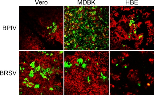
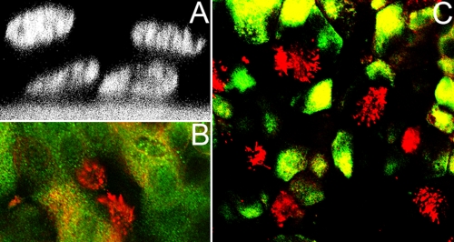
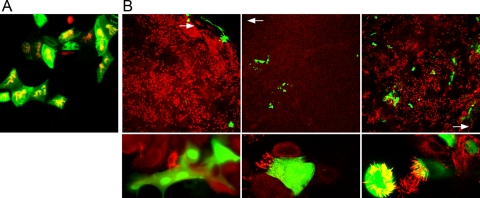
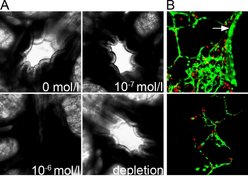
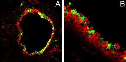
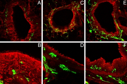
To address the initiation of virus infection in the respiratory tract, we established two culture systems for differentiated bovine airway epithelial cells (BAEC). Filter-grown BAEC differentiated under air-liquid interface (ALI) conditions to generate a pseudo-stratified mucociliary epithelium. Alternatively, precision-cut lung slices (PCLS) from the bovine airways were generated that retained the original composition and distribution of differentiated epithelial cells. With both systems, epithelial cells were readily infected by bovine parainfluenza virus 3 (BPIV3). Ciliated cells were the most prominent cell type affected by BPIV3. Surprisingly, differentiated BAEC were resistant to infection by bovine respiratory syncytial virus (BRSV), when the virus was applied at the same multiplicity of infection that was sufficient for infection by BPIV3. In the case of PCLS, infection by BRSV was observed in cells located in lower cell layers but not in epithelial cells facing the lumen of the airways. The identity of the infected cells could not be determined because of a lack of specific antibodies. Increasing the virus titer 30-fold resulted in infection of the ALI cultures of BAEC, whereas in PCLS the ciliated epithelium was still refractory to infection by BRSV. These results indicate that differentiated BAEC are readily infected by BPIV3 but rather resistant to infection by BRSV. Disease caused by BRSV may require that calves encounter environmental stimuli that render BAEC susceptible to infection.


Figures

FIG. 1.
FIG. 1.
Infection of continuous cell lines (Vero, MDBK, and HBE) by BPIV3 and BRSV. Confluent cells grown on collagenized filter supports were inoculated with virus at an MOI of 0.1 from the apical side. At 2 dpi, cultures were fixed and stained with an antibody for β-tubulin (red). Virus-infected cells are visualized by the expression of GFP (green).
FIG. 2.
FIG. 2.
ALI cultures of BAEC. (A) Pseudostratified epithelium of BAEC grown for 2 weeks under ALI conditions and visualized by DAPI staining and confocal laser scanning microscopy. (B) Mucociliary differentiation of 2-week-old ALI cultures of BAEC. Cilia were stained by an antibody recognizing β-tubulin (red). Mucus-producing cells were detected by an antibody specific for mucin-5AC. (C) Distribution of ciliated cells within the respiratory epithelium. At 2 weeks after ALI culturing, BAEC were stained for cytoceratin (green), an epithelial cell marker, and for β-tubulin to detect cilia (red).
FIG. 3.
FIG. 3.
Well-differentiated BAEC infected by BPIV3 (A) or BRSV (B). (A) BPIV3 was applied to the apical surface at an MOI of 0.1 for 2 h. At 2 dpi, cultures were fixed and cilia were visualized by staining for β-tubulin (red). Virus-infected cells were detected by immunostaining (green). (B) BRSV infection of pseudostratified epithelia of BAEC. Infected cells were fixed 3 (left and right panels) and 10 (middle panels) dpi, respectively, and stained with an antibody recognizing β-tubulin (red). Virus-infected cells were visualized by GFP expression (green). BRSV applied at an MOI of 0.1 (left and middle panels) predominantly infected cells in the border area of the filter, where differentiation was less developed (white arrow, top panels). The infected cells were not ciliated (bottom panels, higher magnification). When virus was applied at an MOI of 3.5 (right panels), BRSV-infected well-differentiated BAEC cells were distributed all over the filter (top panel, lower magnification), including ciliated cells (bottom panel, higher magnification).
FIG. 4.
FIG. 4.
Vitality of PCLS 1 day after preparation. (A) Bronchoconstriction. PCLS prepared from the bronchioli and cultured for 1 day were treated with different concentrations of methacholine. The addition of methacholine at a concentration of 10−6 mol/liter induced bronchoconstriction within seconds (lower left panel). The removal of methacholine resulted in a reversion of this effect (lower right panel). (B) Live/dead viability assay. Live cells are shown in green; dead cells are shown in red. The cells of the respiratory epithelium facing the airway are indicated by a white arrow (top panel). The bottom panel shows alveoli.
FIG. 5.
FIG. 5.
BPIV3 infection of PCLS. At 2 dpi, the slices were stained for virus antigen (green) and β-tubulin (red). Pictures were taken at lower (A) and higher (B) magnifications.
FIG. 6.
FIG. 6.
BRSV infection of PCLS. One day after preparation, PCLS were inoculated with BRSV-GFP. Infected cells are shown in green. For comparison, cells were stained for β-tubulin (red). PCLS infected with 105 PFU/ml were stained at 2 (A), 5 (B) or 10 (C and D) dpi. PCLS infected with 107 PFU/ml were stained at 2 (E) or 5 (F) dpi. For clarity, the images in panels B, D, and F are shown at a higher magnification.

References

    1. Bals, R., C. Beisswenger, S. Blouquit, and T. Chinet. 2004. Isolation and air-liquid interface culture of human large airway and bronchiolar epithelial cells. J. Cyst. Fibros. 249-51. - PubMed
    1. Bose, S., M. Basu, and A. K. Banerjee. 2004. Role of nucleolin in human parainfluenza virus type 3 infection of human lung epithelial cells. J. Virol. 788146-8158. - PMC - PubMed
    1. Chanock, R. M., B. R. Murphy, and P. L. Collins. 2001. Parainfluenza viruses, p. 1341-1380. In D. M. Knipe and P. M. Howley (ed.), Fields virology, 4th ed., vol. 1. Lippincott/The Williams & Wilkins Co., Philadelphia, PA.
    1. Ebsen, M., G. Mogilevski, O. Anhenn, V. Maiworm, D. Theegarten, J. Schwarze, and K. Morgenroth. 2002. Infection of murine precision cut lung slices (PCLS) with respiratory syncytial virus (RSV) and Chlamydophila pneumoniae using the Krumdieck technique. Pathol. Res. Pract. 198747-753. - PubMed
    1. González-Reyes, L., M. B. Ruiz-Argüello, B. Garcia-Barreno, L. Calder, J. A. López, J. P. Albar, J. J. Skehel, D. C. Wiley, and J. A. Melero. 2001. Cleavage of the human respiratory syncytial virus fusion protein at two distinct sites is required for activation of membrane fusion. Proc. Natl. Acad. Sci. USA 989859-9864. - PMC - PubMed

Publication types

MeSH terms

LinkOut - more resources